Martínez JV, Verbanaz SC, Jordán R, Enríquez N, Efrón ED. [Chronic meningococcemia]. Medicina (B Aires). 2008. 68 (4):298-300. [QxMD MEDLINE Link].
Lau C-Y, Tremont EC. Meningococcus and miscellaneous neisseriae. Cunha CB. Schiossberg's Clinical Infectious Disease. 3rd. Oxford University Press; 2020. 946-952.
Welch SB, Nadel S. Treatment of meningococcal infection. Arch Dis Child. 2003 Jul. 88(7):608-14. [QxMD MEDLINE Link]. [Full Text].
Wang X, Theodore MJ, Mair R, Trujillo-Lopez E, du Plessis M, Wolter N, et al. Clinical validation of multiplex real-time PCR assays for detection of bacterial meningitis pathogens. J Clin Microbiol. 2012 Mar. 50 (3):702-8. [QxMD MEDLINE Link].
[Guideline] Crum-Cianflone N. Prevention and control of meningococcal disease: recommendations for use of meningococcal vaccines in pediatric patients. Infect Dis Ther. 2016. 116(2):89-112. [QxMD MEDLINE Link].
[Guideline] Prevention and control of meningococcal disease. MMWR Recomm Rep. 2013 Mar 22. 62:1-22. [QxMD MEDLINE Link].
Perea-Milla E, Olalla J, Sánchez-Cantalejo E, Martos F, Matute-Cruz P, Carmona-López G, et al. Pre-hospital antibiotic treatment and mortality caused by invasive meningococcal disease, adjusting for indication bias. BMC Public Health. 2009 Apr 3. 9:95. [QxMD MEDLINE Link].
Dhanoa RK, Selvaraj R, Shoukrie SI, Zahra A, Malla J, Selvamani TY, et al. Eculizumab's Unintentional Mayhem: A Systematic Review. Cureus. 2022 Jun. 14 (6):e25640. [QxMD MEDLINE Link].
Ladhani SN, Lucidarme J, Parikh SR, Campbell H, Borrow R, Ramsay ME. Meningococcal disease and sexual transmission: urogenital and anorectal infections and invasive disease due to Neisseria meningitidis. Lancet. 2020 Jun 13. 395 (10240):1865-1877. [QxMD MEDLINE Link].
Folaranmi TA, Kretz CB, Kamiya H, MacNeil JR, Whaley MJ, Blain A, et al. Increased Risk for Meningococcal Disease Among Men Who Have Sex With Men in the United States, 2012-2015. Clin Infect Dis. 2017 Sep 1. 65 (5):756-763. [QxMD MEDLINE Link].
Kratz MM, Weiss D, Ridpath A, Zucker JR, Geevarughese A, Rakeman J, et al. Community-Based Outbreak of Neisseria meningitidis Serogroup C Infection in Men who Have Sex with Men, New York City, New York, USA, 2010-2013. Emerg Infect Dis. 2015 Aug. 21 (8):1379-86. [QxMD MEDLINE Link].
Horino T, Kato T, Sato F, Sakamoto M, Nakazawa Y, Yoshida M, et al. Meningococcemia without meningitis in Japan. Intern Med. 2008. 47(17):1543-7. [QxMD MEDLINE Link].
Pollard AJ, Nadel S, Ninis N, Faust SN, Levin M. Emergency management of meningococcal disease: eight years on. Arch Dis Child. 2007 Apr. 92(4):283-6. [QxMD MEDLINE Link]. [Full Text].
Zughaier SM. Neisseria meningitidis capsular polysaccharides induce inflammatory responses via TLR2 and TLR4-MD-2. J Leukoc Biol. 2011 Mar. 89(3):469-80. [QxMD MEDLINE Link]. [Full Text].
Coureuil M, Join-Lambert O, Lécuyer H, Bourdoulous S, Marullo S, Nassif X. Pathogenesis of meningococcemia. Cold Spring Harb Perspect Med. 2013 Jun 1. 3 (6):[QxMD MEDLINE Link].
Brandtzaeg P, van Deuren M. Classification and pathogenesis of meningococcal infections. Methods Mol Biol. 2012. 799:21-35. [QxMD MEDLINE Link].
Brozna JP. Shwartzman reaction. Semin Thromb Hemost. 1990 Oct. 16 (4):326-32. [QxMD MEDLINE Link].
Cimé-Aké E, Carranza-Enríquez F, Hurtado-Arias JJ, Muñoz-Castañeda WRA, Medina-Fonseca B, Barrera-Vargas A, et al. Primary meningococcal septic arthritis associated with joint calcium oxalate crystals: A case report and review of the literature. Mod Rheumatol Case Rep. 2022 Jun 24. 6 (2):296-300. [QxMD MEDLINE Link].
Livorsi DJ, Stenehjem E, Stephens DS. Virulence factors of gram-negative bacteria in sepsis with a focus on Neisseria meningitidis. Contrib Microbiol. 2011. 17:31-47. [QxMD MEDLINE Link].
Plant L, Sundqvist J, Zughaier S, Lovkvist L, Stephens DS, Jonsson AB. Lipooligosaccharide structure contributes to multiple steps in the virulence of Neisseria meningitidis. Infect Immun. 2006 Feb. 74(2):1360-7. [QxMD MEDLINE Link]. [Full Text].
Sanders MS, van Well GT, Ouburg S, Morré SA, van Furth AM. Toll-like receptor 9 polymorphisms are associated with severity variables in a cohort of meningococcal meningitis survivors. BMC Infect Dis. 2012 May 11. 12:112. [QxMD MEDLINE Link].
Pathan N, Faust SN, Levin M. Pathophysiology of meningococcal meningitis and septicaemia. Arch Dis Child. 2003 Jul. 88(7):601-7. [QxMD MEDLINE Link]. [Full Text].
Faust SN, Levin M, Harrison OB, Goldin RD, Lockhart MS, Kondaveeti S, et al. Dysfunction of endothelial protein C activation in severe meningococcal sepsis. N Engl J Med. 2001 Aug 9. 345(6):408-16. [QxMD MEDLINE Link].
Pathan N, Hemingway CA, Alizadeh AA, Stephens AC, Boldrick JC, Oragui EE, et al. Role of interleukin 6 in myocardial dysfunction of meningococcal septic shock. Lancet. 2004 Jan 17. 363(9404):203-9. [QxMD MEDLINE Link].
Pathan N, Williams EJ, Oragui EE, Stephens AC, Levin M. Changes in the interleukin-6/soluble interleukin-6 receptor axis in meningococcal septic shock. Crit Care Med. 2005 Aug. 33(8):1839-44. [QxMD MEDLINE Link].
Bergounioux J, Coureuil M, Belli E, Ly M, Cambillau M, Goudin N, et al. Experimental evidences of human coronary microvasculature and myocardial tissue bacterial colonization during meningococcemia. Infect Immun. 2016 Aug 1. [QxMD MEDLINE Link].
MacLennan J, Kafatos G, Neal K, Andrews N, Cameron JC, Roberts R, et al. Social behavior and meningococcal carriage in British teenagers. Emerg Infect Dis. 2006 Jun. 12(6):950-7. [QxMD MEDLINE Link]. [Full Text].
Andersen UØ, Bay JT, Jensen LH. [Recurrent meningococcal infection in a young woman witha mutation in the C8B gene]. Ugeskr Laeger. 2022 Jun 6. 184 (23):[QxMD MEDLINE Link].
Faber J, Henninger N, Finn A, Zenz W, Zepp F, Knuf M. A toll-like receptor 4 variant is associated with fatal outcome in children with invasive meningococcal disease. Acta Paediatr. 2009 Mar. 98(3):548-52. [QxMD MEDLINE Link].
Jansen AG, Sanders EA, VAN DER Ende A, VAN Loon AM, Hoes AW, Hak E. Invasive pneumococcal and meningococcal disease: association with influenza virus and respiratory syncytial virus activity?. Epidemiol Infect. 2008 Nov. 136(11):1448-54. [QxMD MEDLINE Link]. [Full Text].
Fijen CA, Kuijper EJ, te Bulte MT, Daha MR, Dankert J. Assessment of complement deficiency in patients with meningococcal disease in The Netherlands. Clin Infect Dis. 1999 Jan. 28 (1):98-105. [QxMD MEDLINE Link].
Ladhani SN, Campbell H, Lucidarme J, Gray S, Parikh S, Willerton L, et al. Invasive meningococcal disease in patients with complement deficiencies: a case series (2008-2017). BMC Infect Dis. 2019 Jun 14. 19 (1):522. [QxMD MEDLINE Link].
MacNeil JR, Blain AE, Wang X, Cohn AC. Current Epidemiology and Trends in Meningococcal Disease-United States, 1996-2015. Clin Infect Dis. 2018 Apr 3. 66 (8):1276-1281. [QxMD MEDLINE Link].
Bacterial meningitis. CDC. Available at http://www.cdc.gov/meningitis/bacterial.html. 2015; Accessed: May 2015.
[Guideline] Cohn AC, MacNeil JR, Clark TA, Ortega-Sanchez IR, Briere EZ, Meissner HC, et al. Prevention and control of meningococcal disease: recommendations of the Advisory Committee on Immunization Practices (ACIP). MMWR Recomm Rep. 2013 Mar 22. 62 (RR-2):1-28. [QxMD MEDLINE Link].
Ortega-Sanchez IR, Meltzer MI, Shepard C, Zell E, Messonnier ML, Bilukha O, et al. Economics of an adolescent meningococcal conjugate vaccination catch-up campaign in the United States. Clin Infect Dis. 2008 Jan 1. 46(1):1-13. [QxMD MEDLINE Link].
Cathie K, Levin M, Faust SN. Drug use in acute meningococcal disease. Arch Dis Child Educ Pract Ed. 2008 Oct. 93(5):151-8. [QxMD MEDLINE Link].
Mbaeyi SA, Joseph SJ, Blain A, Wang X, Hariri S, MacNeil JR. Meningococcal Disease Among College-Aged Young Adults: 2014-2016. Pediatrics. 2019 Jan. 143 (1):[QxMD MEDLINE Link].
Centers for Disease Control and Prevention (CDC). Meningococcal Disease, Surveillance Data Tables. Centers for Disease Control and Prevention (CDC). Available at https://www.cdc.gov/meningococcal/surveillance/surveillance-data.html#figure01. Accessed: 2021 Sep 7.
José Francisco Santos N, Viviane Matos F, Caroline Alves F, Martha Silva MS, Leila Carvalho C. Carriage prevalence of Neisseria meningitidis in the Americas in the 21st century: a systematic review. Braz J Infect Dis. 2019 Jul 22. [QxMD MEDLINE Link].
Tappero JW, Reporter R, Wenger JD, Ward BA, Reeves MW, Missbach TS, et al. Meningococcal disease in Los Angeles County, California, and among men in the county jails. N Engl J Med. 1996 Sep 19. 335(12):833-40. [QxMD MEDLINE Link].
Brundage JF, Ryan MA, Feighner BH, Erdtmann FJ. Meningococcal disease among United States military service members in relation to routine uses of vaccines with different serogroup-specific components, 1964-1998. Clin Infect Dis. 2002 Dec 1. 35(11):1376-81. [QxMD MEDLINE Link].
Mandal S, Wu HM, MacNeil JR, Machesky K, Garcia J, Plikaytis BD, et al. Prolonged university outbreak of meningococcal disease associated with a serogroup B strain rarely seen in the United States. Clin Infect Dis. 2013 Aug. 57 (3):344-8. [QxMD MEDLINE Link].
Simon MS, Weiss D, Gulick RM. Invasive meningococcal disease in men who have sex with men. Ann Intern Med. 2013 Aug 20. 159 (4):300-1. [QxMD MEDLINE Link].
Bozio CH, Blain A, MacNeil J, Retchless A, Weil LM, Wang X, et al. Meningococcal Disease Surveillance in Men Who Have Sex with Men - United States, 2015-2016. MMWR Morb Mortal Wkly Rep. 2018 Sep 28. 67 (38):1060-1063. [QxMD MEDLINE Link].
Miller L, Arakaki L, Ramautar A, Bodach S, Braunstein SL, Kennedy J, et al. Elevated risk for invasive meningococcal disease among persons with HIV. Ann Intern Med. 2014 Jan 7. 160 (1):30-7. [QxMD MEDLINE Link].
Sejvar JJ, Johnson D, Popovic T, Miller JM, Downes F, Somsel P, et al. Assessing the risk of laboratory-acquired meningococcal disease. J Clin Microbiol. 2005 Sep. 43 (9):4811-4. [QxMD MEDLINE Link].
Rosenstein NE, Perkins BA, Stephens DS, Lefkowitz L, Cartter ML, Danila R, et al. The changing epidemiology of meningococcal disease in the United States, 1992-1996. J Infect Dis. 1999 Dec. 180 (6):1894-901. [QxMD MEDLINE Link].
Centers for Disease Control and Prevention (CDC). Notice to Healthcare Providers: Recognizing and Reporting Serogroup B Meningococcal Disease Associated with Outbreaks at Princeton University and the University of California at Santa Barbara. Available at http://emergency.cdc.gov/han/han00357.asp. November 27, 2013;
Wilder-Smith A. W135 meningococcal carriage in association with the Hajj pilgrimage 2001: the Singapore experience. Int J Antimicrob Agents. 2003 Feb. 21(2):112-5. [QxMD MEDLINE Link].
Wilder-Smith A, Barkham TM, Earnest A, Paton NI. Acquisition of W135 meningococcal carriage in Hajj pilgrims and transmission to household contacts: prospective study. BMJ. 2002 Aug 17. 325(7360):365-6. [QxMD MEDLINE Link]. [Full Text].
Wilder-Smith A, Chow A, Goh KT. Emergence and disappearance of W135 meningococcal disease. Epidemiol Infect. 2010 Jul. 138(7):976-8. [QxMD MEDLINE Link].
Hart CA, Cuevas LE. Meningococcal disease in Africa. Ann Trop Med Parasitol. 1997 Oct. 91 (7):777-85. [QxMD MEDLINE Link].
Vyse A, Wolter JM, Chen J, Ng T, Soriano-Gabarro M. Meningococcal disease in Asia: an under-recognized public health burden. Epidemiol Infect. 2011 Apr 15. 1-19. [QxMD MEDLINE Link]. [Full Text].
Invasive meningococcal disease in England: annual laboratory confirmed reports for epidemiological year 2017 to 2018. Public Health England. Available at https://assets.publishing.service.gov.uk/government/uploads/system/uploads/attachment_data/file/751821/hpr3818_IMD.pdf. October 26, 2018; Accessed: August 13, 2019.
Sharip A, Sorvillo F, Redelings MD, Mascola L, Wise M, Nguyen DM. Population-based analysis of meningococcal disease mortality in the United States: 1990-2002. Pediatr Infect Dis J. 2006 Mar. 25 (3):191-4. [QxMD MEDLINE Link].
Christensen H, May M, Bowen L, Hickman M, Trotter CL. Meningococcal carriage by age: a systematic review and meta-analysis. Lancet Infect Dis. 2010 Dec. 10(12):853-61. [QxMD MEDLINE Link].
Filippakis D, Gkentzi D, Dimitriou G, Karatza A. Neonatal meningococcal disease: an update. J Matern Fetal Neonatal Med. 2020 Nov 24. 1-6. [QxMD MEDLINE Link].
Moura AS, Pablos-Mendez A, Layton M, Weiss D. Epidemiology of meningococcal disease, New York City, 1989-2000. Emerg Infect Dis. 2003 Mar. 9(3):355-61. [QxMD MEDLINE Link]. [Full Text].
Kaplan SL, Schutze GE, Leake JA, Barson WJ, Halasa NB, Byington CL, et al. Multicenter surveillance of invasive meningococcal infections in children. Pediatrics. 2006 Oct. 118(4):e979-84. [QxMD MEDLINE Link].
Darton T, Guiver M, Naylor S, Jack DL, Kaczmarski EB, Borrow R, et al. Severity of meningococcal disease associated with genomic bacterial load. Clin Infect Dis. 2009 Mar 1. 48(5):587-94. [QxMD MEDLINE Link].
Bouneb R, Mellouli M, Regaieg H, Majdoub S, Chouchène I, Boussarsar M. Meningococcemia complicated by myocarditis in a 16-year-old young man: a case report. Pan Afr Med J. 2018. 29:149. [QxMD MEDLINE Link].
Gawalkar AA, Tale S, Chhabria BA, Bhalla A. Myocarditis and purpura fulminans in meningococcaemia. QJM. 2017 Nov 1. 110 (11):755-756. [QxMD MEDLINE Link].
Zeidan A, Tariq S, Faltas B, Urban M, McGrody K. A case of primary meningococcal pericarditis caused by Neisseria meningitidis serotype Y with rapid evolution into cardiac tamponade. J Gen Intern Med. 2008 Sep. 23(9):1532-5. [QxMD MEDLINE Link]. [Full Text].
Vienne P, Ducos-Galand M, Guiyoule A, Pires R, Giorgini D, Taha MK, et al. The role of particular strains of Neisseria meningitidis in meningococcal arthritis, pericarditis, and pneumonia. Clin Infect Dis. 2003 Dec 15. 37(12):1639-42. [QxMD MEDLINE Link].
Wong JS, Balakrishnan V. Neisseria meningitidis endogenous endophthalmitis: case report and literature review. J Pediatr Ophthalmol Strabismus. 1999 May-Jun. 36(3):145-52. [QxMD MEDLINE Link].
Feldman C, Anderson R. Meningococcal pneumonia: a review. Pneumonia (Nathan). 2019. 11:3. [QxMD MEDLINE Link].
Garcia NS, Castelo JS, Ramos V, Rezende GS, Pereira FE. Frequency of myocarditis in cases of fatal meningococcal infection in children: observations on 31 cases studied at autopsy. Rev Soc Bras Med Trop. 1999 Sep-Oct. 32 (5):517-22. [QxMD MEDLINE Link].
Borg J, Christie D, Coen PG, Booy R, Viner RM. Outcomes of meningococcal disease in adolescence: prospective, matched-cohort study. Pediatrics. 2009 Mar. 123(3):e502-9. [QxMD MEDLINE Link].
Buysse CM, Raat H, Hazelzet JA, Hulst JM, Cransberg K, Hop WC, et al. Long-term health status in childhood survivors of meningococcal septic shock. Arch Pediatr Adolesc Med. 2008 Nov. 162(11):1036-41. [QxMD MEDLINE Link].
Buysse CM, Oranje AP, Zuidema E, Hazelzet JA, Hop WC, Diepstraten AF, et al. Long-term skin scarring and orthopaedic sequelae in survivors of meningococcal septic shock. Arch Dis Child. 2009 May. 94(5):381-6. [QxMD MEDLINE Link].
Huang L, Fievez S, Goguillot M, Marié L, Bénard S, Elkaïm A, et al. A database study of clinical and economic burden of invasive meningococcal disease in France. PLoS One. 2022. 17 (4):e0267786. [QxMD MEDLINE Link].
Stephens DS, Greenwood B, Brandtzaeg P. Epidemic meningitis, meningococcaemia, and Neisseria meningitidis. Lancet. 2007 Jun 30. 369(9580):2196-210. [QxMD MEDLINE Link].
Feldman HA. Meningococcal infections. Adv Intern Med. 1972. 18:117-40. [QxMD MEDLINE Link].
Thimmesch M, Bodart E, Gavage P, Misson JP, Frère J. [Two case reports of meningococcemia. Review of the literature on chronic meningococcemia]. Arch Pediatr. 2016 Jun. 23 (6):595-8. [QxMD MEDLINE Link].
Prista-Leão B, Almeida F, Carvalho AC, Silva S, Sarmento A. Chronic meningococcemia. IDCases. 2019. 15:e00502. [QxMD MEDLINE Link].
Feldman HA. Meningococcal infections. Adv Intern Med. 1972. 18:117-40. [QxMD MEDLINE Link].
Giri S, Shrestha B, Gajurel BP, Sapkota D, Gautam N, Shrestha A. Staphylococcal endocarditis with meningitis and basal ganglia infarcts mimicking meningococcemia. Clin Case Rep. 2022 Mar. 10 (3):e05548. [QxMD MEDLINE Link].
Durand ML, Calderwood SB, Weber DJ, Miller SI, Southwick FS, Caviness VS Jr, et al. Acute bacterial meningitis in adults. A review of 493 episodes. N Engl J Med. 1993 Jan 7. 328 (1):21-8. [QxMD MEDLINE Link].
Parmentier L, Garzoni C, Antille C, Kaiser L, Ninet B, Borradori L. Value of a novel Neisseria meningitidis--specific polymerase chain reaction assay in skin biopsy specimens as a diagnostic tool in chronic meningococcemia. Arch Dermatol. 2008 Jun. 144(6):770-3. [QxMD MEDLINE Link].
Periappuram M, Taylor MR, Keane CT. Rapid detection of meningococci from petechiae in acute meningococcal infection. J Infect. 1995 Nov. 31(3):201-3. [QxMD MEDLINE Link].
Arend SM, Lavrijsen AP, Kuijken I, van der Plas RN, Kuijper EJ. Prospective controlled study of the diagnostic value of skin biopsy in patients with presumed meningococcal disease. Eur J Clin Microbiol Infect Dis. 2006 Oct. 25(10):643-9. [QxMD MEDLINE Link].
Dolan Thomas J, Hatcher CP, Satterfield DA, Theodore MJ, Bach MC, Linscott KB, et al. sodC-based real-time PCR for detection of Neisseria meningitidis. PLoS One. 2011 May 5. 6(5):e19361. [QxMD MEDLINE Link]. [Full Text].
Guarner J, Greer PW, Whitney A, Shieh WJ, Fischer M, White EH, et al. Pathogenesis and diagnosis of human meningococcal disease using immunohistochemical and PCR assays. Am J Clin Pathol. 2004 Nov. 122(5):754-64. [QxMD MEDLINE Link].
Fernandez-Rodriguez A, Alcala B, Alvarez-Lafuente R. Real-time polymerase chain reaction detection of Neisseria meningitidis in formalin-fixed tissues from sudden deaths. Diagn Microbiol Infect Dis. 2008 Apr. 60(4):339-46. [QxMD MEDLINE Link].
Bryant PA, Li HY, Zaia A, Griffith J, Hogg G, Curtis N, et al. Prospective study of a real-time PCR that is highly sensitive, specific, and clinically useful for diagnosis of meningococcal disease in children. J Clin Microbiol. 2004 Jul. 42(7):2919-25. [QxMD MEDLINE Link]. [Full Text].
de Filippis I, do Nascimento CR, Clementino MB, Sereno AB, Rebelo C, Souza NN, et al. Rapid detection of Neisseria meningitidis in cerebrospinal fluid by one-step polymerase chain reaction of the nspA gene. Diagn Microbiol Infect Dis. 2005 Feb. 51(2):85-90. [QxMD MEDLINE Link].
Lin HW, Yin JH, Lo JP, Wang YH, Lee SY, Lu JJ. Use of universal polymerase chain reaction assay and endonuclease digestion for rapid detection of Neisseria meningitides. J Microbiol Immunol Infect. 2004 Dec. 37(6):371-4. [QxMD MEDLINE Link].
Richardson DC, Louie L, Louie M, Simor AE. Evaluation of a rapid PCR assay for diagnosis of meningococcal meningitis. J Clin Microbiol. 2003 Aug. 41(8):3851-3. [QxMD MEDLINE Link]. [Full Text].
Health Protection Agency. Guidance for the public health management of meningococcal disease in the UK. [Full Text].
[Guideline] Updated recommendations for use of meningococcal conjugate vaccines --- Advisory Committee on Immunization Practices (ACIP), 2010. MMWR Morb Mortal Wkly Rep. 2011 Jan 28. 60(3):72-6. [QxMD MEDLINE Link].
van de Beek D, Cabellos C, Dzupova O, Esposito S, Klein M, Kloek AT, et al. ESCMID guideline: diagnosis and treatment of acute bacterial meningitis. Clin Microbiol Infect. 2016 May. 22 Suppl 3:S37-62. [QxMD MEDLINE Link].
Sprung CL, Annane D, Keh D, Moreno R, Singer M, Freivogel K, et al. Hydrocortisone therapy for patients with septic shock. N Engl J Med. 2008 Jan 10. 358(2):111-24. [QxMD MEDLINE Link].
Jaeschke R, Angus DC. Living with uncertainty in the intensive care unit: should patients with sepsis be treated with steroids?. JAMA. 2009 Jun 10. 301(22):2388-90. [QxMD MEDLINE Link].
Brouwer MC, McIntyre P, de Gans J, Prasad K, van de Beek D. Corticosteroids for acute bacterial meningitis. Cochrane Database Syst Rev. 2010 Sep 8. CD004405. [QxMD MEDLINE Link].
Herrera R, Hobar PC, Ginsburg CM. Surgical intervention for the complications of meningococcal-induced purpura fulminans. Pediatr Infect Dis J. 1994 Aug. 13(8):734-7. [QxMD MEDLINE Link].
Besner GE, Klamar JE. Integra Artificial Skin as a useful adjunct in the treatment of purpura fulminans. J Burn Care Rehabil. 1998 Jul-Aug. 19(4):324-9. [QxMD MEDLINE Link].
Faibis S, Widmer R, Sapir S, Peretz B, Shapira J. Meningococcal septicaemia and dental complications: a literature review and two case reports. Int J Paediatr Dent. 2005 May. 15(3):213-9. [QxMD MEDLINE Link].
Dretler AW, Rouphael NG, Stephens DS. Progress toward the global control of Neisseria meningitidis: 21st century vaccines, current guidelines, and challenges for future vaccine development. Hum Vaccin Immunother. 2018 May 4. 14 (5):1146-1160. [QxMD MEDLINE Link].
Brown T. ACIP Updates Meningococcal B Vaccine Recommendations. Medscape. Available at http://www.medscape.com/viewarticle/880617. May 25, 2017; Accessed: June 1, 2017.
[Guideline] Patton ME, Stephens D, Moore K, MacNeil JR. Updated Recommendations for Use of MenB-FHbp Serogroup B Meningococcal Vaccine - Advisory Committee on Immunization Practices, 2016. MMWR Morb Mortal Wkly Rep. 2017 May 19. 66 (19):509-513. [QxMD MEDLINE Link].
Aaberge IS, Oster P, Helland OS, Kristoffersen AC, Ypma E, Høiby EA, et al. Combined administration of meningococcal serogroup B outer membrane vesicle vaccine and conjugated serogroup C vaccine indicated for prevention of meningococcal disease is safe and immunogenic. Clin Diagn Lab Immunol. 2005 May. 12(5):599-605. [QxMD MEDLINE Link]. [Full Text].
Bethell D, Pollard AJ. Meningococcal vaccines. Expert Rev Vaccines. 2002 Jun. 1(1):75-84. [QxMD MEDLINE Link].
Pollard AJ. Global epidemiology of meningococcal disease and vaccine efficacy. Pediatr Infect Dis J. 2004 Dec. 23(12 Suppl):S274-9. [QxMD MEDLINE Link].
Tucker ME. New meningococcal vaccine recommended for high-risk infants. Medscape Medical News. Jan 24, 2013. Available at http://www.medscape.com/viewarticle/778124. Accessed: Feb 5, 2013.
Infant Meningococcal Vaccination: Advisory Committee on Immunization Practices (ACIP) Recommendations and Rationale. MMWR Morb Mortal Wkly Rep. 2013 Jan 25. 62:52-4. [QxMD MEDLINE Link].
Brown T. ACIP OKs Meningitis Vaccine (Menveo) for High-Risk Infants. Medscape Medical News. Available at http://www.medscape.com/viewarticle/813066. Accessed: October 28, 2013.
Basta NE, Mahmoud AA, Wolfson J, Ploss A, Heller BL, Hanna S, et al. Immunogenicity of a Meningococcal B Vaccine during a University Outbreak. N Engl J Med. 2016 Jul 21. 375 (3):220-8. [QxMD MEDLINE Link].
Update: Guillain-Barre syndrome among recipients of Menactra meningococcal conjugate vaccine--United States, June 2005-September 2006. MMWR Morb Mortal Wkly Rep. 2006 Oct 20. 55(41):1120-4. [QxMD MEDLINE Link].
McNamara LA, Potts C, Blain AE, Retchless AC, Reese N, Swint S, et al. Detection of Ciprofloxacin-Resistant, β-Lactamase-Producing Neisseria meningitidis Serogroup Y Isolates - United States, 2019-2020. MMWR Morb Mortal Wkly Rep. 2020 Jun 19. 69 (24):735-739. [QxMD MEDLINE Link].
Fraser A, Gafter-Gvili A, Paul M, Leibovici L. Prophylactic use of antibiotics for prevention of meningococcal infections: systematic review and meta-analysis of randomised trials. Eur J Clin Microbiol Infect Dis. 2005 Mar. 24(3):172-81. [QxMD MEDLINE Link].
Atkinson B, Gandhi A, Balmer P. History of Meningococcal Outbreaks in the United States: Implications for Vaccination and Disease Prevention. Pharmacotherapy. 2016 Aug. 36 (8):880-92. [QxMD MEDLINE Link].
Marshall GS, Dempsey AF, Srivastava A, Isturiz RE. US College Students Are at Increased Risk for Serogroup B Meningococcal Disease. J Pediatric Infect Dis Soc. 2019 May 11. [QxMD MEDLINE Link].
Soeters HM, Whaley M, Alexander-Scott N, Kanadanian KV, MacNeil JR, Martin SW, et al. Meningococcal Carriage Evaluation in Response to a Serogroup B Meningococcal Disease Outbreak and Mass Vaccination Campaign at a College-Rhode Island, 2015-2016. Clin Infect Dis. 2017 Apr 15. 64 (8):1115-1122. [QxMD MEDLINE Link].
Ladhani SN, Giuliani MM, Biolchi A, Pizza M, Beebeejaun K, Lucidarme J, et al. Effectiveness of Meningococcal B Vaccine against Endemic Hypervirulent Neisseria meningitidis W Strain, England. Emerg Infect Dis. 2016 Feb. 22 (2):309-11. [QxMD MEDLINE Link].
Tuncer AM, Gur I, Ertem U, et al. Once daily ceftriaxone for meningococcemia and meningococcal meningitis. Pediatr Infect Dis J. 1988 Oct. 7(10):711-3. [QxMD MEDLINE Link].
Hart CA, Cuevas LE. Meningococcal disease in Africa. Ann Trop Med Parasitol. 1997 Oct. 91(7):777-85. [QxMD MEDLINE Link].
U.S. Food and Drug Administration. First vaccine approved by FDA to prevent serogroup B Meningococcal disease. Available at http://www.fda.gov/NewsEvents/Newsroom/PressAnnouncements/ucm420998.htm. October 29, 2014;
[Guideline] American Academy of Pediatrics Committee on Infectious Diseases. Updated recommendations on the use of meningococcal vaccines. Pediatrics. 2014 Aug. 134 (2):400-3. [QxMD MEDLINE Link].
Harrison LH, Shutt KA, Arnold KE, Stern EJ, Pondo T, Kiehlbauch JA, et al. Meningococcal carriage among Georgia and Maryland high school students. J Infect Dis. 2015 Jun 1. 211 (11):1761-8. [QxMD MEDLINE Link].
Yue M, Xu J, Yu J, Shao Z. Carriage prevalence of Neisseria meningitidis in China, 2005-2022: a systematic review and meta-analysis. BMC Infect Dis. 2022 Jul 7. 22 (1):594. [QxMD MEDLINE Link].
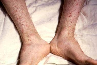
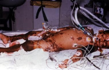
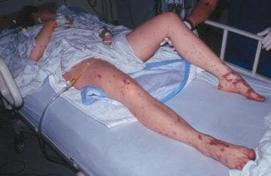 The legs of a 22-year-old woman in septic shock with a rapidly evolving purpuric rash. Photo by D. Scott Smith, MD, taken at Stanford University Hospital.