History
Typically, Stevens-Johnson syndrome (SJS) begins with a nonspecific upper respiratory tract infection. This usually is part of a 1- to 14-day prodrome during which fever, sore throat, chills, headache, and malaise may be present. Vomiting and diarrhea are occasionally noted as part of the prodrome.
Mucocutaneous lesions develop abruptly. Clusters of outbreaks last from 2-4 weeks. The lesions typically are nonpruritic.
A history of fever or localized worsening should suggest a superimposed infection; however, fever has been reported to occur in up to 85% of cases.
Involvement of oral and/or mucous membranes may be severe enough that patients may not be able to eat or drink. Patients with genitourinary involvement may complain of dysuria or an inability to void.
A history of a previous outbreak of Stevens-Johnson syndrome or of erythema multiforme may be elicited. Recurrences may occur if the responsible agent is not eliminated or if the patient is re-exposed.
Typical prodromal symptoms are as follows:
-
Cough productive of a thick purulent sputum
-
Headache
-
Malaise
-
Arthralgia
Patients may complain of a burning rash that begins symmetrically on the face and the upper part of the torso. This may be accompanied by ocular symptoms.
In addition to the skin, lesions in Stevens-Johnson syndrome may involve the following parts of the body:
-
Oral mucosa
-
Esophagus
-
Pharynx
-
Larynx
-
Anus
-
Trachea
-
Vagina
-
Urethra
Ocular symptoms include the following:
-
Red eye
-
Tearing
-
Dry eye
-
Pain
-
Blepharospasm
-
Itching
-
Grittiness
-
Heavy eyelid
-
Foreign body sensation
-
Decreased vision
-
Burning sensation
-
Photophobia
-
Diplopia
Delineation of a drug exposure timeline is essential, especially in the 1-3 weeks preceding the cutaneous eruption.
Physical Examination
The rash can begin as macules that develop into papules, vesicles, bullae, urticarial plaques, or confluent erythema. The center of these lesions may be vesicular, purpuric, or necrotic.
The typical lesion has the appearance of a target; this is considered pathognomonic. However, in contrast to the typical lesions of erythema multiforme, these lesions have only two zones of color. The core may be vesicular, purpuric, or necrotic; that zone is surrounded by macular erythema. Some have called these targetoid lesions.
Lesions may become bullous and later rupture, leaving denuded skin. The skin becomes susceptible to secondary infection. Extensive sloughing is shown in the image below.
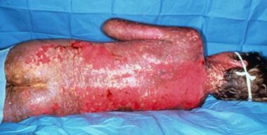 Note extensive sloughing of epidermis from Stevens-Johnson syndrome. Courtesy of David F. Butler, MD.
Urticarial lesions typically are not pruritic. Infection may be responsible for the scarring associated with morbidity.
Although lesions may occur anywhere, the palms, soles, dorsum of the hands, and extensor surfaces are most commonly affected. Desquamation on the foot is shown in the image below.
 Sheetlike desquamation on the foot in a patient with toxic epidermal necrolysis. Courtesy of Robert Schwartz, MD.
The rash may be confined to any one area of the body, most often the trunk.
Mucosal involvement may include erythema, edema, sloughing, blistering, ulceration, and necrosis. An example of this type of involvement is shown in the image below.
 Hemorrhagic crusting of the mucous membranes in toxic epidermal necrolysis. Similar lesions are seen in Stevens-Johnson syndrome. Courtesy of Robert Schwartz, MD.
For more information, see the Medscape Reference article Dermatologic Manifestations of Stevens-Johnson Syndrome and Toxic Epidermal Necrolysis.
Although some have suggested the possibility of Stevens-Johnson syndrome without skin lesions, most believe that mucosal lesions alone are not enough to establish the diagnosis. Cases without skin lesions have been termed "atypical" or "incomplete." [17] These authors suggested that the combination of urethritis, conjunctivitis, and stomatitis established the diagnosis of Stevens-Johnson syndrome in a patient with Mycoplasma pneumonia– induced signs and symptoms.
The following signs may be noted on examination:
-
Fever
-
Orthostasis
-
Tachycardia
-
Hypotension
-
Altered level of consciousness
-
Epistaxis
-
Conjunctivitis
-
Corneal ulcerations
-
Erosive vulvovaginitis or balanitis
-
Seizures
-
Coma
The following signs may be noted on external examination:
-
Conjunctival hyperemia (ie, red eye)
-
Entropion
-
Skin lesions
-
Nasal lesions
-
Mouth lesions
-
Discharge (ie, catarrhal, mucous, membranous)
The following ocular signs may be noted on slit lamp examination (see the images below):
-
Eyelids: Trichiasis, distichiasis, meibomian gland dysfunction, blepharitis
-
Conjunctiva: Papillae, follicles, keratinization, subepithelial fibrosis, conjunctival shrinkage, foreshortening of fornices, symblepharon, ankyloblepharon
-
Cornea: Superficial punctate keratitis, epithelial defect, stromal ulcer, neovascularization, keratinization, limbitis, conjunctivalization, stromal opacity, perforation
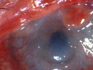 A patient with severe eye involvement associated with Stevens-Johnson syndrome. Note corneal neovascularization and conjunctivalization of the ocular surface.
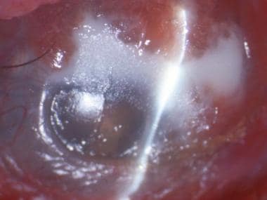 Epithelial defect of the cornea with neovascularization and surface conjunctivalization.


Complications
Of patients with Stevens-Johnson syndrome, 27-50% progress to severe ocular disease. Ocular complications of Stevens-Johnson syndrome include the following:
-
Chronic cicatrizing conjunctivitis
-
Chronic dry eye disease
-
Corneal epithelial defects
-
Corneal stromal ulcers
-
Corneal perforation
-
Endophthalmitis
Other complications may include the following:
-
Gastroenterologic - Esophageal strictures
-
Genitourinary - Renal tubular necrosis, renal failure, penile scarring, vaginal stenosis
-
Pulmonary - Tracheobronchial shedding with resultant respiratory failure
-
Cutaneous - Scarring and cosmetic deformity, recurrences of infection through slow-healing ulcerations
Lesions may continue to erupt in crops for as long as 2-3 weeks. Mucosal pseudomembrane formation may lead to mucosal scarring and loss of function of the involved organ system. Esophageal strictures may occur when extensive involvement of the esophagus exists. Mucosal shedding in the tracheobronchial tree may lead to respiratory failure.
Blindness may develop secondary to severe keratitis or panophthalmitis in 3-10% of patients. Vaginal stenosis and penile scarring have been reported. Renal complications are rare.
Cutaneous lesions may resolve with a patchwork of hyperpigmentation and hypopigmentation. Fingernails and toenails may regrow abnormally. Lesions of the genitourinary system may lead to phimosis or vaginal synechiae.
-
A patient with severe eye involvement associated with Stevens-Johnson syndrome. Note corneal neovascularization and conjunctivalization of the ocular surface.
-
Epithelial defect of the cornea with neovascularization and surface conjunctivalization.
-
Note extensive sloughing of epidermis from Stevens-Johnson syndrome. Courtesy of David F. Butler, MD.
-
Sheetlike desquamation on the foot in a patient with toxic epidermal necrolysis. Courtesy of Robert Schwartz, MD.
-
Hemorrhagic crusting of the mucous membranes in toxic epidermal necrolysis. Similar lesions are seen in Stevens-Johnson syndrome. Courtesy of Robert Schwartz, MD.
-
Note early cutaneous slough with areas of violaceous erythema.
-
Extensive sloughing on the face.
-
Note the presence of both 2-zoned atypical targetoid lesions and bullae.
-
Extensive blistering and sloughing on the back.
-
Extensive sloughing on the back.
-
Note extensive sloughing.
-
Low-power view showing full-thickness epidermal necrosis.


