1/7
The central vein sign on fluid‑attenuating inversion recovery* (FLAIR*) MRI.
2/7
The central vein sign on fluid‑attenuating inversion recovery* (FLAIR*) MRI.
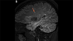
3/7
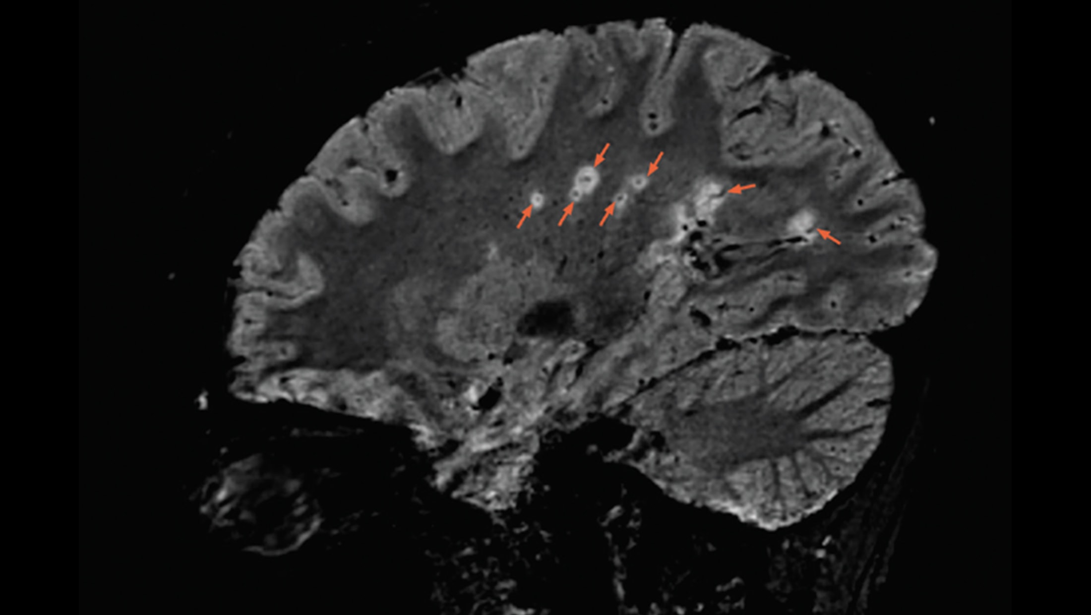
A sagittal 3T FLAIR* brain MRI shows multiple lesions with the central vein sign (orange arrows), appearing as hypointense dots or lines, located centrally within the lesion, depending on the blood vessel orientation. The FLAIR* technique was developed by Daniel S. Reich, MD, PhD and colleagues as described in Sati P, et al. Radiology. 2012;265(3):926-932. Image contributed by Daniel S. Reich, MD, PhD.
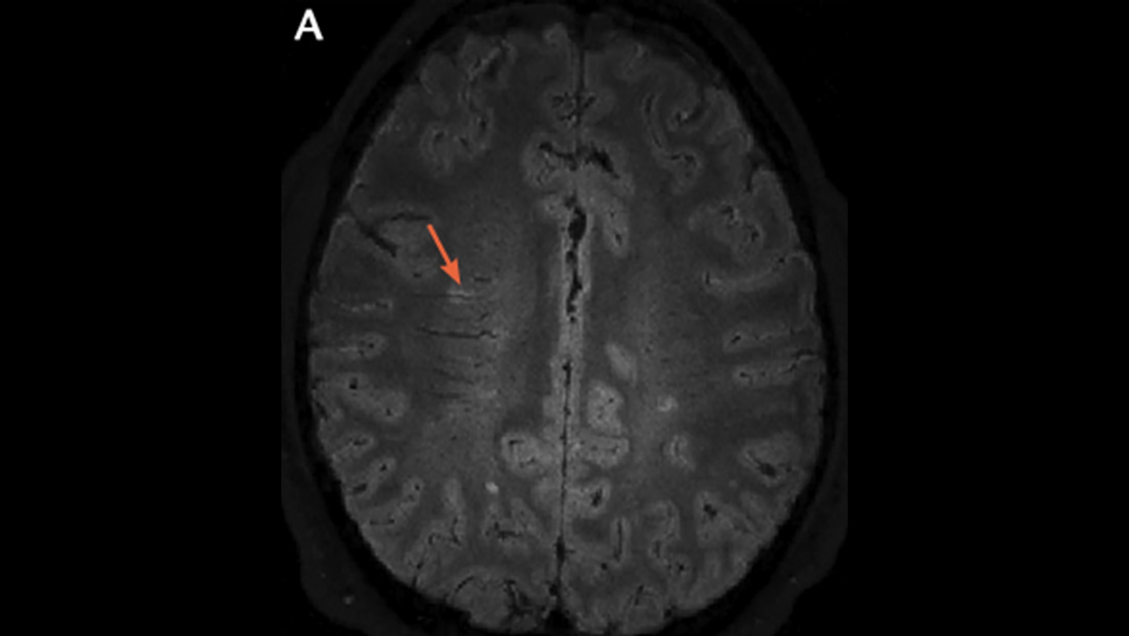
4/7
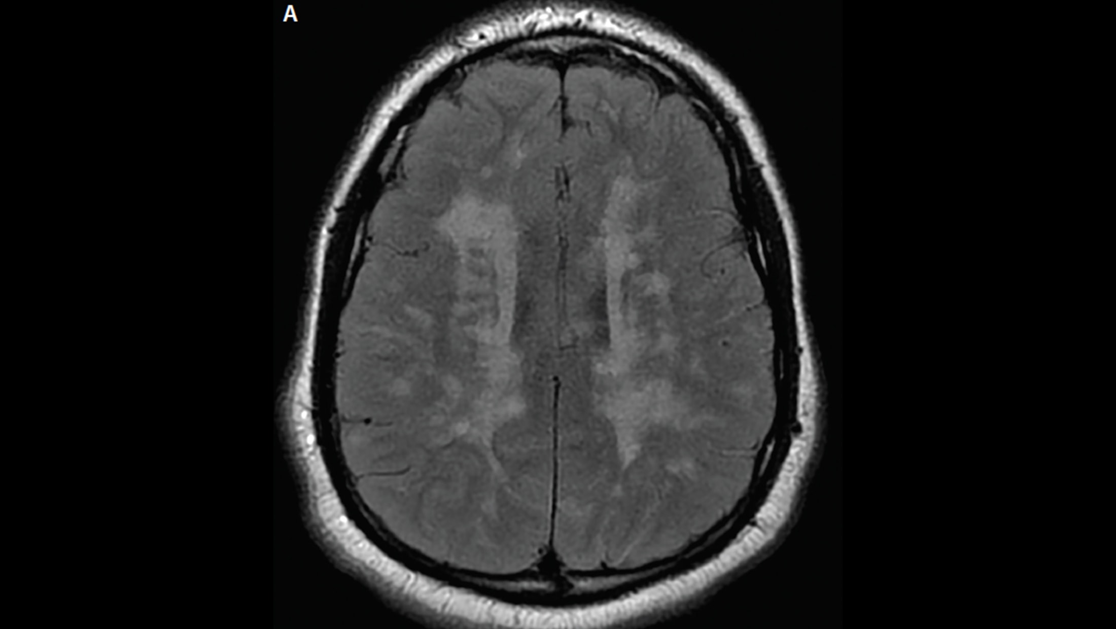
Axial fluid-attenuated inversion recovery (FLAIR) MRI of the brain from a boy, age 16 years, with multiple sclerosis. Note the high lesion burden and areas of early confluence. Children with MS are subject to more frequent relapses and typically have a higher lesion burden than adults with MS. (Images courtesy of Dr. Aaron Carlson.)
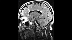
5/7
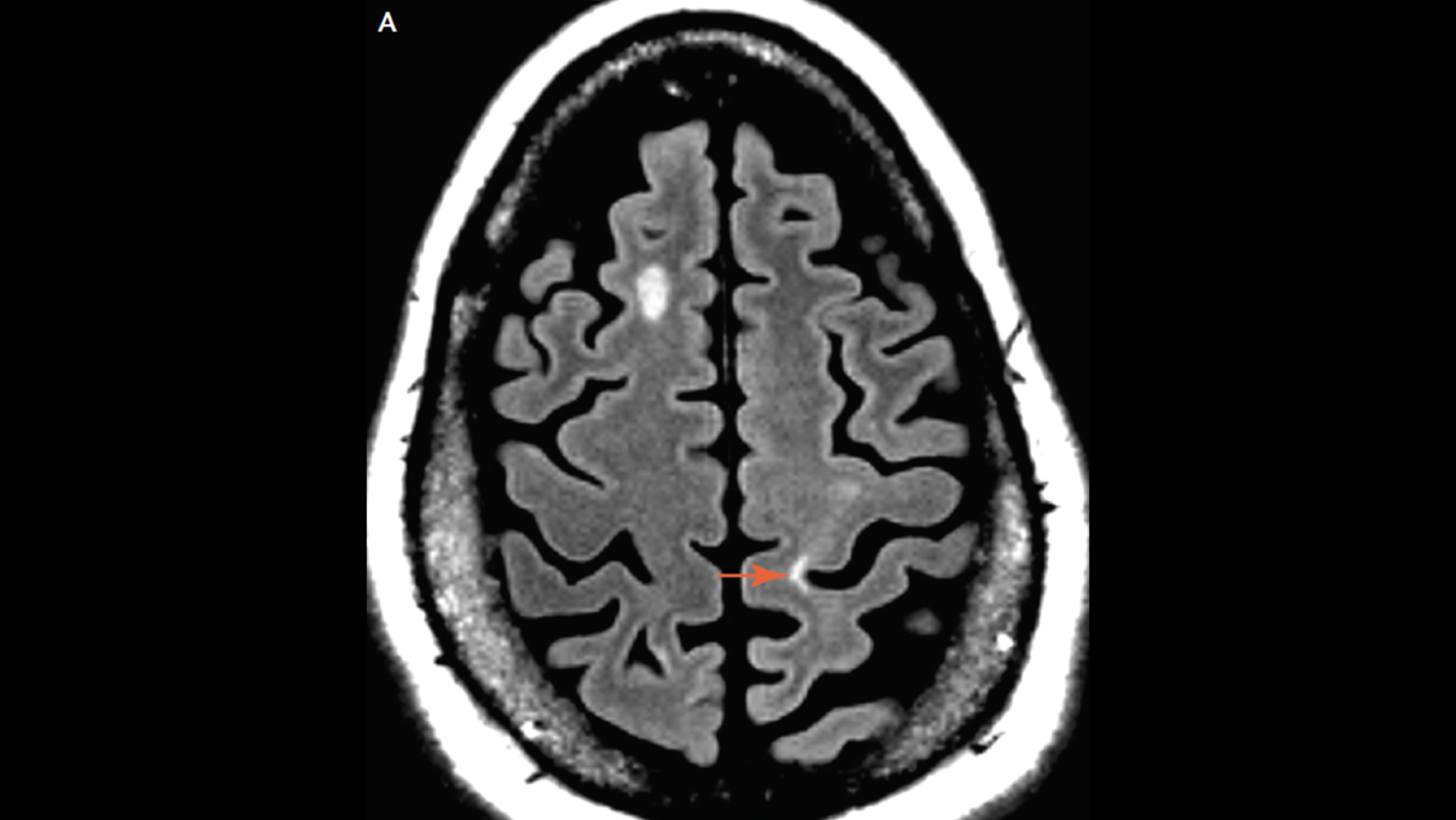
Typical lesions of multiple sclerosis are found in the juxtacortical.
6/7
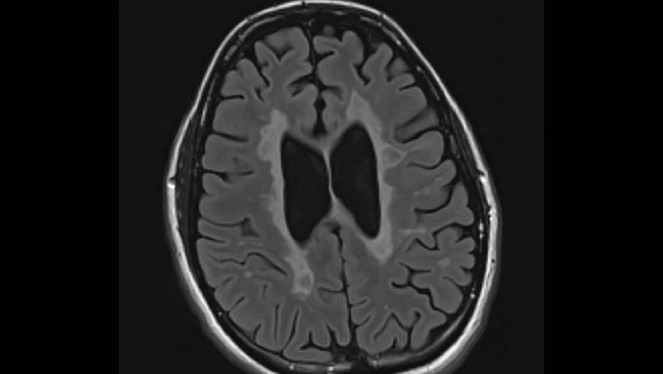
Confluent lesions may appear later in the course of multiple sclerosis.
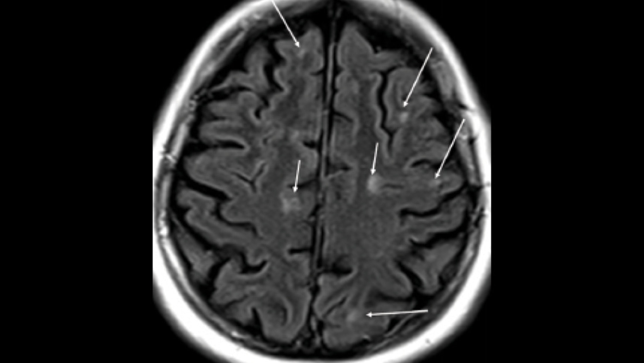
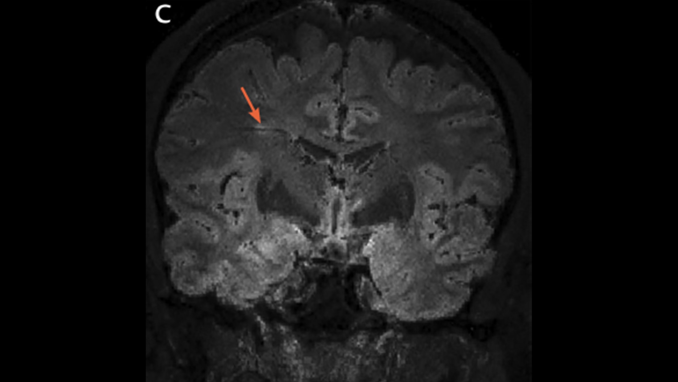
7/7
Cortical, juxtacortical lesions, and U-fiber lesions. Arrows: multiple small juxtacortical and cortical lesions throughout cerebral hemispheres. By definition, no white matter may interpose between a juxtacortical lesion and the cortex. Note U-fiber lesions along arcuate fibers in middle left frontal lobe, highly characteristic of demyelination and not seen in normal aging or vascular disease.