The transmission electron micrographs included in this collection were all the work of Dr. Eldon Newcomb. Dr. Newcomb was a pioneer in the fixing and preparation of plant tissue for viewing with the transmission electron microscope. Dr. Newcomb generously allowed me to scan a sampling of his glass plates on a flat bed scanner. I do not have documentation about the source of the tissue scanned. However, each of these examples illustrates good generic views of plant cellular ultrastructure.
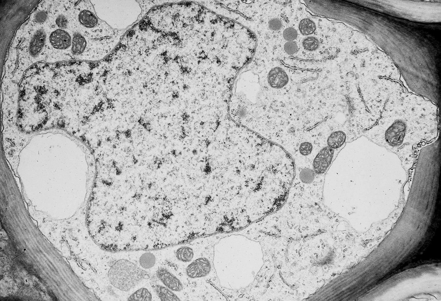
Young plant cells typically have more than one vacuole. These typically merge to form the huge central vacuole of a mature parenchyma cell.