Review

Biofilm Formation, and Related Impacts on Healthcare, Food Processing and Packaging, Industrial Manufacturing, Marine Industries, and Sanitation–A Review

by
Ghazal Shineh
1,
Mohammadmahdi Mobaraki
2,
Mohammad Jabed Perves Bappy
3 and
David K. Mills
4,*
1
School of Biomedical Engineering, University of Sydney, Sydney, NSW 2006, Australia
2
Biomaterial Group, Faculty of Biomedical Engineering (Center of Excellence), Amirkabir University of Technology, Tehran 1591634311, Iran
3
Micro and Nanoscale Systems Engineering, Louisiana Tech University, Ruston, LA 71272, USA
4
School of Biological Sciences, Louisiana Tech University, Ruston, LA 71272, USA
*
Author to whom correspondence should be addressed.
Appl. Microbiol. 2023, 3(3), 629-665; https://doi.org/10.3390/applmicrobiol3030044
Submission received: 16 May 2023 / Revised: 9 June 2023 / Accepted: 22 June 2023 / Published: 26 June 2023

Abstract

:
Biofilm formation can lead to problems in healthcare, water distribution systems, food processing and packaging, industrial manufacturing, marine industries, and sanitation. These microbial communities can proliferate on biotic or abiotic surfaces, and are responsible for human disease and decreasing production efficiency and service equipment life in many industrial fields. The formation of biofilm starts with the attachment of bacteria to the surface, followed by bacterial proliferation and maturation of the microbial community. After forming a biofilm, bacteria not resistant to antimicrobial agents in their planktonic forms can turn resistant. The antibiotic resistance of bacterial biofilm, and the association of biofilms in generating infectious diseases in humans, highlight the need for designing novel and successful antibacterial, anti-biofilm, or anti-infection materials. This paper aims to review the mechanism of biofilm formation, the impact on different industries, the interaction mechanism of nanoparticles with bacteria, and strategies to design anti-biofilm materials. Examples of designing anti-infection bio-implants, coatings, medical devices, wound dressings, and sutures are reviewed.

1. Introduction

Biofilms are complex structures and communities of microorganisms embedded in an extracellular polymeric substance (EPS) matrix and show high antibiotic resistance. The EPS, which is responsible for the cohesion of microorganisms and adhesion of biofilms to surfaces, is composed of proteins including enzymes, DNA, RNA, polysaccharides, and water [1,2]. Both Gram-positive and Gram-negative bacteria can form biofilms. However, the most common biofilm-forming bacteria are Staphylococcus aureus (S. aureus), Enterococcus faecalis, Streptococcus viridans, Staphylococcus epidermidis, Escherichia coli (E. coli), Proteus, Klebsiella pneumoniae, mirabilis, Burkholderia cenocepacia, Clostridium difficile, and Pseudomonas aeruginosa [3,4].
As biofilms can be formed on living or non-living surfaces, they are prevalent in biological, natural, industrial, and hospital settings. Moreover, understanding the role of biofilm in certain infectious diseases, and the inherent tolerance and resistance to antimicrobial treatments, are essential for public health. Hence, biofilm generation has become a significant problem in healthcare, water distribution systems, food processing and packaging, industrial manufacturing, marine industries, and sanitation. Due to its massive impact, extensive research has been performed to understand the biofilm process and its resistance to antibacterial agents [5,6,7,8,9].
With the emergence of drug-resistant bacteria, healthcare-acquired infections have become a major human health problem, as they increase care costs and prolonged hospital stays. In addition, biofilms can lead to a variety of human diseases, such as native valve endocarditis, cystic fibrosis, periodontitis, otitis media, rhinosinusitis, osteomyelitis, chronic bacterial prostatitis, non-healing infected chronic wounds, kidney infections, contact lens-associated keratitis, meningitis, chronic sinusitis, and implantable device-related infections (IDRI) such as prosthetic joint, urinary, and intravascular stent infection [10,11,12,13,14,15].
IDRI can be the most common cause of healthcare-associated infections and severe morbidity and mortality. IDRI is associated with fully implanted devices and those that cross the skin or mucosal barriers. The infection rate in implantable devices relates to the implantation site and intended lifespan [16,17]. For example, dialysis catheter-related bloodstream infection ranges from 3.8 to 5.5 per 1000 catheter days. While for non-dialysis catheters, the infection risk ranges from 2.5 to 4 per 1000 catheters per day.
Aside from the effect of biofilm and microbial contamination on human health, their formation in industrial or marine fields can impose expenses, decrease equipment service life, and even cause human health problems. Hence, designing anti-biofilm materials and substrates for any specific application is essential.

2. Microbial Contamination, Biofilm Formation, and Healthcare-Related Impacts

Microbial contamination is a severe issue, and refers to introducing infectious microorganisms or their toxins. It can lead to many health problems in medical and non-medical fields. The aggregation of microbial cells will cause the formation of biofilm in which bacteria display an exceptional resistance to environmental stresses, especially antibiotics [18]. This section will explain the role of material surface, environmental factors, and the conditioning film on biofilm formation and the mature biofilm structure. Next, the stages of biofilm formation will be explained comprehensively.

2.1. The Role of Material Surface on the Formation of Conditioning Films and Biofilms

Conditioning film is a thin layer consisting of organic and inorganic substances that develops on a material’s surface upon contact with water or other liquids [19]. It comprises proteins, polysaccharides, lipids, and minerals, which can be sourced from the surrounding environment or released by microorganisms. The formation of a conditioning film happens relatively quickly, typically within minutes to hours, and is influenced by factors such as surface characteristics, water chemistry, and the presence of organic components [20].
The presence of a conditioning film is a crucial prerequisite for the formation of biofilms [21,22]. A conditioning film establishes the initial surface for microorganisms to attach to, and creates a favourable environment that promotes the development of biofilms [22].
The conditioning film enhances microbial adhesion via creating microenvironments or introducing chemical cues [23]. This film acts as a reservoir of nutrients for microorganisms. It accumulates organic and inorganic compounds from the surrounding environment, or it is produced by microorganisms within the film. These nutrients serve as a food source for the attached microorganisms, supporting their initial growth and enabling the establishment of a biofilm. This chemical communication promotes the coordinated attachment and growth of microorganisms, facilitating the formation of multicellular communities within the biofilm structure. Additionally, the conditioning film reduces hydrodynamic forces, stabilizing the initial microbial attachment and preventing detachment from the surface [20].
Hence, the formation of a conditioning film leads to changes in the physicochemical properties of the surface after the adsorption of (macro)molecules on the substrate [20], which facilitates the biofilm formation [23].
The formation of conditioning film and biofilm layer on a substrate strongly depends on surface properties, including roughness, hydrophobicity/hydrophilicity, surface charge, stiffness, and surface composition [20,24].
Rough surfaces with a porous structure are highly prone to the formation of conditioning films [20]. The pores allow for the accumulation and retention of organic and inorganic substances, creating an environment conducive to biofilm development [25]. Moreover, the roughness of a surface can provide more areas for bacteria to attach and establish a biofilm [26]. Rough surfaces with crevices and irregularities offer a favourable condition for bacterial adhesion, while smoother surfaces offer fewer attachment sites [25].
The hydrophobic or hydrophilic nature of a surface affects conditioning film formation and bacterial attachment [27]. Surfaces with a high affinity for water, known as hydrophilic surfaces, are more likely to develop conditioning films. The hydrophilic nature promotes the adsorption of water and dissolved substances, including organic compounds, proteins, and polysaccharides, which contribute to the formation of the conditioning film. However, it worth mentioning that a group of bacteria prefer hydrophobic surfaces, while others favour hydrophilic surfaces [28].
The surface charge of a material influences bacterial adhesion [18]. Surfaces with a charge, either positive or negative, exhibit an affinity for conditioning film formation. The presence of charges attracts oppositely charged molecules, such as ions, proteins, and microbial cells, facilitating their accumulation and attachment to the surface. Bacterial cell surfaces also carry a net charge, and the surface charge of the material can affect electrostatic interactions with bacterial cells. Opposite charges can promote adhesion, while similar charges can repel bacterial attachment.
The chemical composition of a surface can also impact biofilm formation. Different materials may have varying affinities for bacterial adhesion. Some materials possess inherent antibacterial properties, which can inhibit or reduce biofilm formation. For example, silver or copper surfaces exhibit antibacterial effects [29].

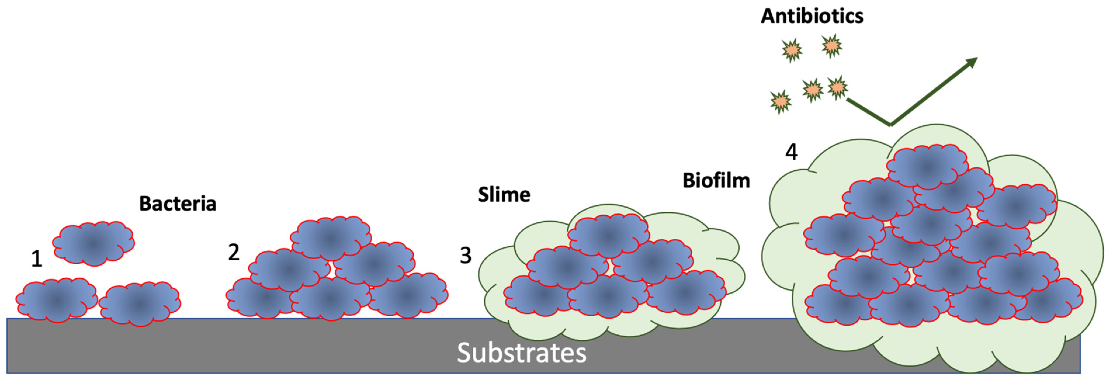
2.2. Biofilm Formation Steps and Its Characteristics

After the formation of the conditioning layer, biofilm development starts via the adhesion of bacteria. Figure 1 depicts biofilm development that typically involves five stages. The first stage is cell attachment to a supportive surface. This stage is followed by the second stage, which is related to cell–cell adhesion and formation of a microcolony. The third stage is cell proliferation, and is followed by slime formation, cell proliferation and, finally, biofilm maturation as four stages. The mature biofilm provides an environment that shields resident bacteria from antibiotics. The fifth or last stage of the biofilm life cycle is bacterial dispersal, which is facilitated by mature biofilm [30].
The first step of biofilm formation, bacterial adhesion, is considered reversible, and is the process where planktonic bacteria move close to the material surface. The initial attachment of 127 bacteria is facilitated by van der Waals interactions and non-covalent forces, and 128 is strengthened by a structural adhesins part of the cellular envelope, such as pilus (plural pili) and 129 flagellum (plural flagella) [31]. Pili and flagella, as bacterial surface appendages, are found on the surface of some bacterial cells as filamentous proteinaceous structures, and perform crucial actions in the adhesion, motility, uptake, and excretion of proteins [32]. Surface characterizations of the material, aside from environmental factors such as pH and temperature, control the bacterial adhesion. For example, bacteria have a lower propensity to be attached to highly hydrophobic surfaces with a water contact angle of more than 90 degrees [33,34]. This is because biomolecules, such as proteins, prefer to be attached to hydrophilic surfaces with high energy. Hence, the conditioning film and consequent biofilm formation chance will be higher on hydrophilic surfaces.
After attachment, the cells start to replicate, microcolonies are developed, and exopolymeric substances (EPS) secretion leads to an irreversible adhesion [35]. The EPS accelerates the proliferation of the adherent bacteria by acting as a scaffold for stabilizing the three-dimensional biofilm structure. As shown in Figure 2, the EPS encloses the E. coli biofilm bacteria, holds microorganisms, and serves as a physical scaffold for the cellular organization [36]. Environmental signals, such as temperature and nutrient availability, encourage the development of bacterial proliferation through quorum sensing (QS). QS plays a vital role in biofilm maturation, and responds to extracellular signalling molecules called autoinducers [37]. Bacteria communicate within and between species through the production and release of autoinducers—the concentration of autoinducers increases in response to a population increase of quorum-sensing bacteria. Hence, autoinducers allow bacteria to sense and monitor their local population density [4]. The bacterial population will alter gene expression and mount coordinated responses to their environments when the intracellular concentration of the autoinducer reaches the minimal threshold.
Nevertheless, gene expression through QS and autoinducers secretion in response to environmental events, such as depletion or availability of nutrients and fluctuations in cell-population density, control the biofilm dispersion [38].
It is worth noting that aside from autoinducers, bacteria residing within biofilms utilize nanowires to communicate with their surroundings and enable bacteria to transmit signals across the biofilm and to neighbouring cells [39,40,41]. While signal molecules are involved in quorum sensing, nanowires play a distinct role in transmitting and receiving signals from the external environment [42]. Bacterial nanowires serve as extensions that enable bacteria to sense and respond to changes in their surrounding environment. They can detect variations in nutrient gradients, redox potential, pH levels, and other environmental parameters. This sensory capability empowers bacteria to adapt their behaviour and coordinate collective responses within the biofilm [41].
After the maturation of the biofilm due to the proliferation of microorganisms, dispersion, the last phase of the biofilm lifecycle starts [43,44]. In this stage, sessile cells start to leave the colony, in order to repeat the cycle by contaminating other surfaces [45].
Bacteria within the biofilm exhibit phenotypes distinct from planktonic bacteria [46], making biofilms a significant public health problem. Biofilm-associated microorganisms gain resistance to immune defence and antibiotics [47,48,49,50,51,52,53,54] and, in comparison to planktonic cultures, they can be up to 1000 times more tolerant to antibiotics [10,55]. It is now well understood the manner in which bacterial biofilms are responsible for up to 75% of human infectious diseases [56]. Intrinsic antimicrobial resistance of biofilms is due to three main reasons. The first reason stems from the fact that EPSs resist the diffusion of antimicrobial agents, through chemically reacting with them or by limiting their transport rate [36,57]. Hence, antimicrobial agents will not be able to contact microorganisms and inactivate them. The second reason is related to the microbial inactivation kinetics. By minimizing their growth rate, biofilm-associated organisms decrease the rate of antimicrobial agents taken into the cell. Bacteria in biofilms are surrounded by an extracellular matrix that physically restricts the diffusion of antimicrobial agents. Nutrient and oxygen depletion within the biofilm causes some bacteria to enter a nongrowing state and initiate microbial inactivation. Third, the environment protects the organisms by providing more protective conditions [58].
A mature biofilm benefits from a favourable living environment for resident bacterial cells and plays a crucial role in maintaining the architecture of the biofilm [59]. The EPS matrix, secreted by microorganisms during growth, comprises polysaccharides, proteins, lipids, nucleic acids, enzymes, and other compounds, such as humic acids [60,61]. Figure 3 represents the biofilm structure comprising the cell cluster, streamer, void, pores, and water channels [62]. The clusters of microbial cells in biofilm create open areas for water flow in the EPS matrix [63,64]. Water constitutes a significant part of a biofilm (up to 97%), and the water flow channels distribute nutrients to microcolonies [1,5,10,65,66].
Aside from bacterial biofilms, fungal biofilms are intricate communities of fungi that adhere to surfaces and form multicellular structures within a matrix of extracellular substances [67]. These biofilms consist of diverse fungal species, each playing a role in the overall structure and function of the biofilm. Fungal biofilms also exhibit distinct characteristics and behaviours compared to individual, free-floating fungi such as resistance to antifungal agents [67].
It is worth noting that fungal biofilms have emerged as significant entities within mixed microbial communities. Their coexistence and interactions with bacteria generate intricate and multifaceted relationships within these communities [68]. These interactions exhibit a dynamic nature, contributing to the complexity of the microbial consortia. For example, fungal biofilms can serve as platforms for bacterial attachment, promoting the formation and stability of mixed biofilms. However, interactions between fungi and bacteria within biofilms can be competitive as well. For example, fungi may hinder biofilm formation by producing antifungal compounds or competing with bacteria for resources and space. Moreover, fungi can also have significant implications for antibiotic resistance in bacteria, as they can form physical barriers that impede antibiotic penetration and diminish their efficacy.
Apart from fungus, the interaction of a virus (bacteriophages) and biofilm can also have implications for biofilm formation and development. Bacteriophages can be used as disinfectant to hinder biofilm formation [69]. However, in the context of antibiotic resistance, bacteriophages can facilitate the transfer of antibiotic resistance genes between bacterial cells through a process called transduction, contributing to the spread of resistance traits within the biofilm [70].
By growing the biofilm on living tissues, medical devices, industrial equipment, and the natural environment, the biofilm microbial community facilitates human disease transmission and increases the risk of persistent disease [6]. It has been believed that the transmission and persistence of human disease can be caused by biofilm formation [6,7].
Figure 1. Graphic illustration of bacterial adhesion mediated by cell surface structures (pili, flagella, fimbriae, and membrane proteins), (1); leading to colonization with cell proliferation, slime (EPS) formation, (2); increased EPS formation, (3); quorum sensing leading to biofilm maturation, (4); formation of microcolonies, and later bacterial dispersal.
Applmicrobiol 03 00044 g001
Figure 2. Scanning electron microscopy image of E. coli biofilm on Teflon. The E. coli bacteria in the biofilm are coloured in pink, and the extracellular matrix is pointed by arrows [3]. Reproduced with permission from [71], 2017, University of Mohammed Premier Oujda.
Applmicrobiol 03 00044 g002
Figure 3. Schematic representation of biofilm architecture. Reproduced with permission from reference [72], 2013, Elsevier.
Applmicrobiol 03 00044 g003

3. Biofilm Formation Challenges to Healthcare, Water Distribution Systems, Food Processing and Packaging, and Marine Industries

3.1. Healthcare

Biofilm growth on living tissues, bioimplants, and external medical devices, generates microbial and chronic infections in the body. They can lead to chronic infections due to their resistance to antibacterial agents and the host immune system. Moreover, their detachment can increase production of emboli and infection in the bloodstream or urinary tract [7].
Bacterial biofilms are responsible for most of the chronic human body infections. However, it is difficult to calculate the real financial impact of biofilms, as many sectors are affected by them. For example, it has been estimated that urinary tract infections, infective endocarditis, and wound infection cost 1, 16, and 281 billion dollars globally per year, respectively [73].
Biofilm-related infections can be categorized as device-related and non-device-related infections. Examples of device-related infections include prosthetic joint infections [74], cardiac implantable electronic device-related infections [75], catheter-associated urinary tract infections [76], and ventilator-associated pneumonia [77], while non-device-related infections include chronic wound infections, urinary tract infections [78], infectious kidney stones [79], periodontitis [80], dental plaque, infective endocarditis [81], and cystic fibrosis [82]. In the human body, microorganisms can colonize the surface of the teeth, skin, and mucosa [83]. Hence, biofilms can be found in the mouth, skin, digestive, respiratory, and reproductive tracts.
Oral microorganisms can be formed on natural teeth, restorative materials, dental implants, and prosthetic constructions [5]. Biofilms are responsible for oral diseases such as dental caries and periodontitis. A sticky film of bacteria, known as plaque, causes cavities and gingivitis by producing acids [6,7]. Dental caries infection can spread to deeper tissues near the root and bloodstream [8].
The colonisation of microorganisms on human skin contributed to certain skin diseases and delayed wound healing [84]. For example, Figure 4 represents acne vulgaris caused by biofilm formation of Cutibacterium acnes (C. acnes) within the follicles, the sebaceous gland, and the pore itself [84,85]. Additionally, S. aureus colonisation can be found in patients with atopic eczema [86]. Furthermore, by producing destructive enzymes and toxins, biofilms can also affect the healing potential of chronic wounds and prolonged hospitalization for some patients [87].
Biofilm formation and infections are the most frequent wound complication, and a major clinical challenge in wound management. The wound healing cascade comprises four overlapping well-orchestrated, integrated phases, including hemostasis, inflammation, proliferation, and remodelling (or maturation).
The first stage of wound healing, or hemostasis, starts once the blood leaks out of the body. In this stage, clots are formed and blood vessels are constricted to prevent blood loss. Next, the inflammation stage involves the participation of inflammatory cells (white blood cells) in the wound area to prevent infection. Next, in the proliferation phase, new granulation tissue is formed, and the wound is contracted, filled, and covered. In the final phase, remodelling, the formed tissue matures into a scar and becomes more robust, elastic, and flexible.
Non-healing or “stalled” wounds result from interruption or interference in these pathways [88]. Wounds will not generally heal if one of the above phases has been disrupted. For instance, infected wounds are stuck in the inflammation or proliferation phases [89]. Hence, wound dressing capable of releasing antibacterial agents in a controlled way can play an essential role in preventing biofilm formation and treating wounds.
Generally, a biofilm-based disease occurs due to two reasons. First, they can result from some impairment of the host defence systems, e.g., chronic lung infection with P. aeruginosa in cystic fibrosis patients [90]. Second, they can be the consequence of the presence of some external or implantable medical device [91]. When a device is exposed to biological fluids, such as blood, urine, and saliva, the surface of the device is rapidly adsorbed with a layer of “conditioning film” [22,23] and initiates the biofilm development. Biofilm formation on indwelling medical devices, such as artificial veins and joints, central venous catheters, prosthetic heart valves, contact lenses, urinary catheters, and intrauterine devices, can lead to serious recalcitrant infections [92]. For instance, the infection rate can be as high as 100% in ureteral catheters and 28% in ureteral stents [93].
Considering the complexity of the interaction of biofilm mechanisms with antibiotic activity, understanding the interplay between critical items, including the immune system, environmental conditions of biofilm infections, phenotypic and genetic resistance mechanisms of biofilms, is required to optimize the antibiotic treatment of biofilm infections [94].
New approaches have been developed to prevent biofilm formation on medical devices. These approaches include the application of antimicrobial coatings, alternating surface topography [95], and incorporation of antibacterial agents (e.g., silver, ciprofloxacin a, fluoroquinolone) [96]. For example, researchers have proved that the surface of nickel-titanium (NiTi) graphene oxide/silver nanoparticles (NPs) can modify medical device (GO/AgNPs) coatings to reduce the consequences of biofilm formation [97]. In another study, it has been proven that a coating containing varied-shaped or non-spherical gold NPs is highly effective against biofilm formation on the surface of latex urinary catheters [98]. In implanted blood-contacting medical devices, combining fluorocarbon chemistry and submicron topography modification in textured crosslinkable octafluoropentoxy phosphazene (X–OFP) materials can reduce the risk of microbial infection [99]. For dental implant application, Nagay et al. [100], via the plasma electrolytic oxidation (PEO), co-opted TiO2 coating with nitrogen and bismuth to achieve an antibacterial effect. More details on this will be given in the following sections.

3.2. Water Distribution Systems

Biofilm formation on pipe walls in the water distribution system is responsible for various water quality, health, operational, and environmental problems. For example, bacteria within these films can affect the water’s turbidity, odour, and taste [54]. Hence, they can exert a chlorine demand. Additionally, the growth of pathogenic bacteria in drinking water distribution systems can cause significant human health risks [9]. Furthermore, bacterial growth can also increase the corrosion of pipe materials. Consequently, drinking water quality will be decreased even more by releasing pipe material and contaminants into the water [101]. Furthermore, these bacteria can also increase the proliferation of some organisms such as Asellus spp. by acting as a trophic food web [102].
One of the critical factors that can encourage or prevent biofilm formation is the type of pipe material [103]. It has been proven that biofilm formation is encouraged on PVC and polyethylene (PE) plastic materials. At the same time, glass is an inert surface and prevents biofilm formation [104]. Hallam et al. [105] demonstrated that biofilm activity is influenced by pipe material far less than chlorine, and biofilm growth potential can be ranked in the order glass < cement < PE < PVC. Ren et al. [106] studied biofilm samples from different pipe materials, and revealed that biofilm morphology and bacterial communities are diverse among different pipe materials.
Surface characteristics of the pipes, such as “biological affinity”, roughness, and surface energy, can control the attachment of bacteria. Therefore, it is needed to test the potential of pipe materials to promote biofilm growth and formation [107]. Another determinant factor is the water flow, with its chemical and hydraulic characteristics [107]. Researchers investigated the combined impact of pipe material and flow hydrodynamics on biofilm development in drinking water distribution systems. They proved that biofilms formed in lower flows had a more significant amount of microbial biomass, and opportunistic pathogens had limited ability to propagate under high shear conditions without roughness [108]. Another study shows that turbulence can enhance the initial formation and development of thick biofilms, due to the increased transport of nutrients and oxygen to the biofilm surface [109]. Water distribution systems are vulnerable to causing health problems for consumers, such as gastrointestinal illness (GII), diarrheal and respiratory diseases, nervous system problems, chronic diseases such as cancer, and adverse effects on reproductive health [110,111,112,113].
Chemical disinfection, particularly with chlorine, is the primary strategy to control biofilm formation in water distribution systems. Due to its ease of use, high solubility, stability, and low cost, chlorine oxidizing agent is a solid and effective disinfectant and prevents microbial regrowth. However, high chlorine concentrations can cause organoleptic problems and increase the production of carcinogens. Other disinfectants besides chlorine, such as chloramines, chlorine dioxide, ozone, and UV radiation, can be used to disinfect drinking water [114].

3.3. Food Processing and Packaging

The food industry is also an example where biofilms can cause economic loss and severe health risks [115]. Bacterial biofilm can be formed on food matrixes, packaging, or factory equipment. Biofilm formations on food manufacturing surfaces will increase the corrosion of the metallic surfaces and can generate unpleasant odours and tastes. Therefore, biofilm will not only cause financial losses, but will also damage the reputations of food companies.
Figure 5 depicts the complex process of biofilm formation on food-contact surfaces. First, a conditioning film is formed by depositing organic molecules on the equipment’s surface. Second, this conditioning film encourages the attachment of biologically active microorganisms. Third, persistent microorganisms remain and initiate growth, even after cleaning and sanitizing. Last, more extensive biofilms are formed [116].
These biofilms may contain pathogenic and toxigenic bacteria, such as Escherichia coli, Listeria monocytogenes, Campylobacter jejuni, Salmonella enterica, Pseudomonas aeruginosa, Bacillus cereus, and S. aureus [117]. Hence, a significant challenge in the food sector is to develop an efficient disinfection strategy. For instance, to eliminate or prevent biofilm formation on the surfaces of the factory equipment, food companies rely on different cost-effective or expensive biofilm removal methods. These methods include physical removal (with hot water steam), chemical treatment (with quaternary ammonium compounds, sodium hydroxide or sodium hypochlorite solutions, or ozone), mechanical removal, non-thermal plasma treatments, enzymatic disruption, application of bacteriophages, and surface modifications [118].
In food packaging, applying many thermal and drying bacteria destruction methods can decrease the food quality [119]. Therefore, new food preservation methods safe for human health and food quality preservation have been developed, such as the production of antibacterial food packaging films and coatings [120]. It has been proven that the shelf life of the vegetables can be increased by aloe vera gel and polyvinyl alcohol (PVA)-based film incorporated with antifungal and antibacterial agents [121]. In addition, bioactive packaging film containing sonorensin, an antimicrobial peptide, showed growth inhibition of spoilage bacteria in chicken meat and tomato samples [122].

3.4. Industrial Manufacturing

Every year, biofilm formation causes an immense loss of industrial productivity and human health-related issues. Microbially induced corrosion brings billions of dollars in losses to US industries annually [123]. The common sectors affected by biofouling and microbial growths include, but are not limited to, power, textiles, apparel manufacturing, chemical, oil, gas production, transportation, shipping, and construction industries. Textile fibres are prone to be affected by microbial growth due to their inherent molecular properties. In apparel manufacturing industries, biofilm growth can cause foul smells, faded colours of the cloths, and even deterioration of the quality of the fabric [124]. The warm and moist environment of the knitting and dyeing floor aggravates the formation of various microorganisms such as Staphylococcus aureus, Escherichia coli, and Pseudomonas sp. [125]. Fungi and moulds can grow very fast on natural fibre-made clothes, as they contain many nutrients for biofilms. Although synthetic fibre-made clothes are less sensitive to microbial growth for their poor absorbing capacity, moistures can be trapped in between the mesh of the fibres and are affected by fungi and bacteria [124]. In addition, cellulolytic bacteria can create cellulolytic enzymes on cellulose fibres and degrade their quality and longevity [126]. Thus, it is a big challenge for the textile industries to minimize biofilm formation. Some biocide compounds, such as chlorinated phenols and organo-coppers, can be applied to reduce the textile materials’ microbial growth by blocking the biofilm cells’ energy production.
Nanotechnology-based solutions are prevalent nowadays to treat fabric. For example, enhanced titanium dioxide (TiO2)-based nanoparticles can be used to catch the dirt and pollutants of the textile material and break the chemical structure into carbon dioxide (CO2) and water (H2O) [127]. In another study, applications of anti-fouling magnetic nanoparticles proved to be another effective method in preventing biofilm development in textile dressing [128].
Manufacturing industries are mainly constructed with concrete and cement-based structures. Microbial-induced concrete corrosion causes massive financial losses in the construction industries. The primary deterioration of the concrete structures causes by a colourless sulphur bacterium named Thiobacillus sp. This microorganism reacts with the sulphur-based compounds and produces sulphuric acid. The produced sulphuric acid reacts with the free lime of the concrete and cement materials and creates various composites of gypsum and calcium. Hence, an internal pressure will be developed inside the concrete, which eventually forms microcracks [129].
It has been proven that acid-producing Fusarium sp. black biofilm (Figure 6) can result in concrete weight and thickness loss [130]. Figure 6B shows a concrete specimen that has been exposed to Fusarium sp. in a humidity chamber for one year. The contaminated specimen showed pH level reduction from 11 to 8 and 2.27 ± 0.16 mm thickness reduction after one year. Decreasing the concrete pH can increase its deterioration rate. Hence, designing and producing antibacterial concrete can prevent bacterial growth and consequent deterioration and biodegradation. For example, it has been shown that applications of ZnO and lignin to concrete can inactivate fungal and bacterial biofilm formation [130].
Nanotechnology-enhanced silicon dioxide, titanium dioxide, zinc peroxide, ferric oxide, and aluminium oxide are incorporated into the concrete to increase its mechanical properties and hydration rate [131]. In addition, titanium dioxide and calcium carbonate nanoparticles can be mixed with fly ash to increase concrete’s durability and anti-bacterial properties [132].
Manufacturing industries also suffer from biofilm growth in the water and gas pipelines, condenser tubes, and storage tanks. Biofilm formation becomes a primary concern for shipping, transportation, energy, power, and chemical companies. Biofilms in water pipelines that are formed by the bacteria present in the water can decrease the flow rate of the line. The formation of bacteria and fungi inside a condenser tube decreases the heat transfer rate and condensation efficiency [133]. As a result, the gas and fuel pipelines, reservoirs, storage tanks, and refining facilities in the oil and gas industries are corroded easily by the attack of sulphate-reducing bacteria [134].
The deterioration can cause pipeline leakage and disastrous incidents in the mentioned fields and industries. Various surface modification techniques are being used to overcome this problem. For instance, electroless plating is a popular method to modify the surface of pipes or tubes to resist biofilm formation. It has been proven that a thin coating of antibacterial copper nanofilm on top of a titanium surface provides a good resistance against biofilm for industrial application [135]. Chemical vapor deposition and physical vapor deposition on the metallic surfaces are also effective but relatively expensive surface modification methods. Additionally, an antimicrobial bilayer of nickel and copper nanofilm can be used on top of a metallic surface to increase its antibacterial properties [136].

3.5. Marine Industries

On surfaces immersed in seawater, the undesirable accumulation of marine organisms, plants, and animals, can lead to the formation of marine biofouling, a natural detrimental phenomenon [137]. The root causes of biofouling are the attachment of microorganisms to the surfaces and bacterial biofilm formation [3]. According to Figure 7, after its attachment, the conditioned film will develop and encourage the adhesion of larger organisms such as algae, mussels, and barnacles to the surface [138]. Marine fouling organisms can be fixed on the surfaces of ships, buoys, offshore installations, and submarine hulls, and on the factory and power station refrigerating equipment surfaces that employ seawater for cooling purposes [139]. As shown in Figure 7, based on the size of the fouling organisms, they can be divided into two groups: (1) microfouling or biofilm, and (2) macrofouling [140].
Additional damage can be generated by biofouling in marine-based industries and infrastructure [141]. For example, as seen in Figure 8, in ship hulls, it will lead to deterioration of the protective coatings, increasing the frequency of dry-docking operations, increasing the fuel consumption, and reducing speed due to the roughness generated by them. The more the fuel consumption, the more the CO2 emissions and transportation costs [142]. Therefore, biofouling can cause both environmental and economic problems [143]. When fixed on the surfaces of buoys, mines, offshore platforms, underwater acoustic devices, cables, and port installations or offshore platforms, their presence will accelerate corrosion processes, increase the materials repair frequency, and reduce operativity. They can also interrupt the electric current generation in power stations [139]. The aforementioned adverse effects have led to the development of various strategies to prevent biofilm formation, such as the application of non-toxic and eco-friendly marine antifouling biolubricant [144], coatings [145,146,147,148,149], and paints [150,151]. Researchers have shown that as an environmentally friendly alternative for antifouling prevention, ultraviolet light (UV) also can be used [152].

3.6. Sanitation

Sanitation against biofilm and microorganisms has recently become a critical concern, especially after the COVID-19 pandemic. Some Gram-positive bacteria, such as methicillin-resistant Staphylococcus aureus (MRSA) and vancomycin-resistant enterococci (VRE), can survive from days to months on the surface of fabrics and plastics [153]. It has been shown that 65% of the nurses who are involved in treating patients with MRSA bacteria wounds can contaminate their garments with MRSA bacteria [154]. Although MRSA is the leading cause of difficult-to-treat infections, and remains a concern in hospitals, the importance of sanitizers increased after the recent pandemic caused by COVID-19.
When it comes to the food industry and food service preparation area, sanitation plays an important role, since it can directly affect the wellbeing and health of people. Hence, effective sanitation methods can reduce the risk of pathogens spreading.
Sanitization can be performed mainly in two ways: chemical sanitation and physical sanitation. Chemical sanitation includes the application of agents such as chlorine and ammonium compounds, acids, peroxides, and silver compounds. For example, chlorine-based sanitizers are widely accepted for their cheap cost, disinfecting power, and oxidizing capabilities [155]. The efficacy of the sanitizer depends on the pH level, temperature, contact time, and presence of other unwanted organic compounds, such as dirt, soil, etc. [156]. Chemical sanitizers are often mixed with other cleaning agents, such as detergents or enzymes, to increase efficacy [157,158].
An investigation with several active chemical compounds’ performance in the sanitization of Staphylococcus aureus, Proteus mirabilis, and Pseudomonas aeruginosa biofilms is shown in Table 1.
Recently, alcohol-based chemical compounds have been widely used to resist bacterial growth on human skin, plastic, and rubber. A mixture of ethanol and isopropanol or n-propanol is often used to sanitize medical instruments, hospital areas, offices, and building floors [171]. Alcohol solutions can damage the cell structure and protein-membrane of the bacteria, and proved to be an excellent disinfectant against pathogens [172]. It has been proven that the WHO-recommended formulation hand antiseptic, which is alcohol-based, is capable of killing 99.90% of bacteria [173].
Aside from chemical-based sanitation methods, physical sanitization techniques are also effective in removing biofilms. The huge scope of industrial applications is the reason behind the recent popularity of this method. The major of types of physical sanitation techniques are based on high temperatures, radiation, and plasma. High temperature-based sanitation methods, or thermal disinfection, is a universal and effective sanitation technique. Dry or wet heat can be applied to remove biofilm and kill bacteria by denaturing their proteins. Steam sterilisation via autoclave is an example of thermal disinfection.
Plasma, as the fourth state of matter, contains electrons and ions. These unstable charged particles (radicals) can kill or inactivate pathogens via reacting with nucleic acids, proteins, and lipids. Plasma-based sanitation techniques are also widely used in medical, food agriculture, and environmental fields [174]. For example, in the medical and dentistry filed, plasma techniques can not only be used in medical device sterilization, but can also be used in tooth and root canal disinfection and pathogen-based skin diseases [174]. Cleaning of food processing, packaging and distribution equipment is an example of the plasma disinfecting technique in the food industry [175]. In the agriculture industry, not only are plasma methods applied to disinfect fertilizers, seeds, soil, and water, but they can also enhance plant growth, through increasing antioxidative activity via short-lived radicals. In the environmental field, plasma techniques are used in wastewater cleaning, and treatment of exhaust gases.
Atmospheric pressure glow discharges (APGD) are effective for both biofilm and planktonic microorganisms. In this method, highly reactive atomic oxygen, ozone, or hydroxyl radicals are discharged in high atmospheric pressure to sterilize microorganisms [175]. A combined method of biofilm removal by sonication and proteolytic enzymes also can very effectively remove Escherichia coli from a surface [176]. Ultrasound enhancement procedures can increase the performance of biocides for the control of biofouling [177]. The efficacy of biocides and antibiotics can also be increased by using an electric enhancement to kill biofilm bacteria. Experiments show that biofilm bacteria can readily be killed when biocides are placed on the area of an active electrode [178].

3.7. Biofilm in Microbiologically Influenced Corrosion (MIC) of Metallic Alloys in the Oil and Gas Industry

MIC poses a significant challenge in the oil and gas industry, resulting in substantial financial losses [179]. The metallic alloys Inconel and Monel are widely used in the oil and gas industry due to their exceptional resistance to corrosion. However, when exposed to microorganisms, these alloys can still undergo corrosion, specifically galvanic corrosion and surface pitting [180,181,182,183]. Galvanic corrosion, also known as bimetallic corrosion, arises when two dissimilar metals or alloys come into contact in the presence of an electrolyte. It occurs due to the difference in electrical potentials between the metals, leading to accelerated corrosion of the more reactive metal. Surface pitting refers to the localized corrosion attack on a metal or alloy surface, resulting in the formation of small pits or cavities. It can occur due to various factors, including aggressive ions, localized cell concentrations, and corrosive environments.
The presence of bacteria and biofilms can exacerbate galvanic corrosion by influencing the local electrochemical conditions [181]. The presence of diverse bacteria with distinct electrochemical properties can lead to the formation of galvanic cells on the surface of the alloy. This occurs when different bacteria create microenvironments with varying oxygen concentrations or release corrosive by-products, which disrupt the electrochemical balance and accelerate corrosion [184].
In addition, biofilms can create microenvironments with variations in oxygen availability, pH, and ion concentration. Bacterial biofilms can generate localized variations in oxygen concentration on the alloy surface, forming oxygen-concentration cells. In these cells, areas with low oxygen levels serve as anodic sites, while areas with higher oxygen levels act as cathodic sites. This creates a galvanic couple, intensifying corrosion and promoting the formation of surface pits [185].
In addition, bacteria present within the biofilm produce metabolic by-products, such as organic acids, which create localized corrosive conditions. These by-products lower the pH near the alloy surface, elevate the concentration of aggressive materials such as sulphuric acid, and facilitate the generation of corrosive substances, all of which contribute to surface pitting [186,187].
Understanding the intricate interactions between bacteria, metallic alloys, and the formation of galvanic elements within the alloy vacancies, is crucial for effectively addressing and mitigating MIC in the oil and gas industry. Implementing strategies to control biofilm formation, manage microbial metabolism, and modify the electrochemical conditions can help minimize the occurrence and impact of corrosion on metallic alloys.

4. Recent Trends in Antimicrobial Material Research, Infection Control and Prevention

Antibiotic-resistant bacterial infections impose substantial healthcare problems and lead to higher morbidity and mortality. Treating antibiotic-resistant bacteria infection with currently available antibiotics is more complicated and, in some instances, impossible [188,189]. Designing an antibacterial, antifouling, anti-biofilm, or anti-infection surfaces and materials decreases the need for systemic antibiotic therapy and the chance of antibiotic resistance developing in various bacterial species. According to Figure 9, in this section, the strategies to develop anti-biofilm surfaces, together with some examples, will be introduced. Then, methods to develop anti-biofilm bulk materials will be reviewed. In the end, nanoparticle-bacteria interactions, and mechanisms by which NPs kill bacteria cells, will be further explained.

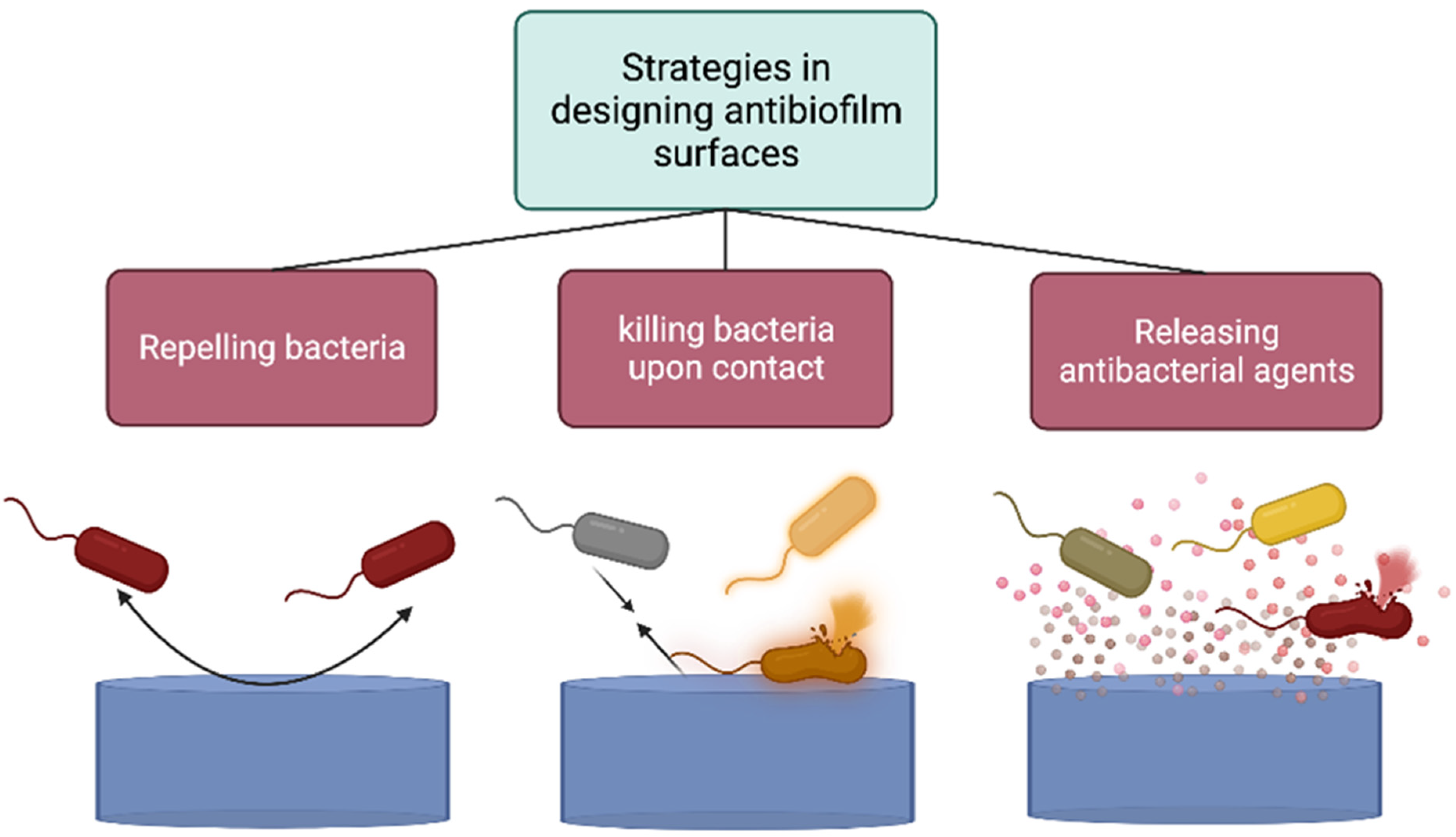
4.1. Strategies to Design Anti-Biofilm or Antifouling Surfaces and Materials

Engineered surfaces capable of reducing the adhesion, proliferation, and spread of bacteria can prevent infections, reduce the use of antibiotics, and limit disinfectants (Figure 9). Learning from nature, natural antifouling surfaces appear to use a combination of physical structure and chemical composition to inhibit biofouling [190].
Marine sessile organisms, such as fishes, seaweeds, and jellyfish, cannot adhere to living organisms. In contrast, they can adhere to hard solid surfaces such as a ship’s hull, rocks, and concrete walls [191]. For example, from the view of surface characteristics, researchers proved that surface roughness and structure play an important role in providing natural antifouling properties. Furthermore, considering the chemical composition and biomimicking of sea organisms, several hundred natural antifouling chemical compounds have been discovered [191,192]. A schematic depiction of strategies to prevent the formation of biofilm is shown in Figure 9. According to Figure 10, to design an antimicrobial material, inspired by nature, there are two main biomimetic strategies, namely: (1) designing protein repelling, cell repelling, or self-cleaning surface, and (2) designing bactericidal surfaces [193].

4.2. Designing Protein-Repellent, Cell-Repellent, and Self-Cleaning Surfaces

The first strategy to provide antibacterial and anti-biofilm material is to prevent the attachment of bacteria by designing protein repellent or self-cleaning surfaces. As can be seen in Figure 10, such surfaces can be provided by designing bio-mimicked unfavourable surface topography and structure, by changing the chemical composition of the surface through anti-biofouling polymers, or by the immobilization of anti-foulant groups.

4.2.1. Mimicking the Surface Microtopography of Marine Organisms

Mimicking the surface microtopography of marine organisms encourages researchers to design unfavourable surface topography for microorganisms and design anti-biofilm surfaces for a variety of applications. For instance, micro-structured riblets found on the dermal denticles of shark skin provide self-cleaning and antifouling properties, as depicted in Figure 11. The fouling resistance of Polymethyl methacrylate (PMMA) shark skin patterns and of the smooth controls against biofilm growth were evaluated using Staphylococcus aureus and Escherichia coli as the model bacteria. The smooth surface structure and nanostructured protuberances are possessed by the ridges, riblets and grooved surface, respectively [193]. The denticles comprised a regular pattern of parallel ridges and grooves, and the combination of the denticle’s flexibility, together with its surface texture, also led to a reduction in microbial attachment [193,194].
On soft PMMA surfaces made by topographies mimicking shark skin, a 67% reduction in fouling was observed [195]. Considering the surface structures of shark skin, gecko feet, butterfly wings, and lotus leaves, a strong relationship between nano-structure surfaces, self-cleaning, and even bactericidal effects have been discovered [195]. Based on marine organisms’ surface microtopographies, several key surface parameters can generate anti-biofouling surfaces. These parameters include lower values of mean surface roughness, a high skewness of roughness and waviness, a low fractal dimension, and higher values of anisotropy [192,196,197].

4.2.2. Covering the Surface of Solids with Antifouling Polymers or the Chemical Immobilization of Anti-Foulant Groups

Another method to repel bacteria and prevent biofilm formation is to cover the surfaces of solids with antifouling polymers, or to immobilize chemical anti-foulant groups on the surfaces, as illustrated in Figure 10. For example, grafting the surface with hydrophilic polymers, such as polyethylene glycol (PEG) [198,199,200,201], zwitterionic superhydrophilicity polymers [202,203,204], or even superhydrophobic antifouling polymers, prevents biofilm formation by reducing protein adsorption and subsequently cells [204]. Researchers grafted a telomer of dodecafluoroheptyl methacrylate (DFMA), triclosan acrylate (TA), and 3-mercaptopropyl trimethoxysilane (KH590) to bis-silanol terminated poly(dimethylsiloxane) (PDMS). They proved that this coating inhibits the growth of marine bacterial biofilm after immersion in natural seawater for more than six months [205]. Researchers immobilized polyzwitterions onto silicon surfaces to develop an antifouling silicon surface for implantable medical devices, such as artificial kidney devices [206]. Wang et al. [186] prepared new types of zwitterionic polymer network (ZPN) coatings that have self-healing, excellent protein repellence, and anti-biofouling properties. The antimicrobial effectiveness of the mechanism involves the selective binding of the zwitterionic polymers to oppositely charged species through electrostatic interactions. Furthermore, any scratching on these coatings can be repaired in water. Moreover, after healing, both the anti-biofouling and mechanical properties can be repaired [207].
The healing mechanism in such coatings relies on the interaction between negative and positive charges, and the reformation of broken ionic bonds in the presence of water [207]. Liu et al. [145] designed a self-repairing silicone-polymer-based coating that can effectively inhibit biofouling or release biofoulants for marine applications. The mechanical properties of these coatings could be recovered in either the air or in artificial seawater at room temperature, and accelerated at a higher temperature. In a study, researchers developed a dual-function coating with antifouling and self-healing properties that was coated on a 316 L stainless steel substrate with a solid adhesive strength [208]. The anti-protein adhesion efficiency of the coating reached 90%, and could inhibit the adhesion of biofoulants due to the presence of hydroxyethyl acrylate (HEA) and poly (ethylene glycol) methyl ether acrylate (PEGA480) units. Moreover, the coating could be repaired within 15 min after being scratched, and the mechanical property was restored to 93% of its original condition [188]. Nguyen et al. [209] proved that zwitterionic poly (sulfobetaine acrylamide) brushes grafted from silicon-rich silicon nitride surfaces exhibit excellent protein repellency (>99%) during exposure to a fibrinogen (FIB) solution. Co-deposition of fluorinated particles during electroless nickel plating represents an effective and commercially scalable method to prepare antifouling coatings on stainless steel for food processing [210].
Coating materials incorporated with fluoroalkylated acrylic acid oligomer (FAAO) were used to coat resin composite substrates to design a surface with the self-cleaning property. When the concentration of FAAO increased, the surface of the coating materials possessed self-cleaning properties and inhibited biofilm formation [211]. Acosta et al. [212] immobilized antimicrobial peptides (AMPs) on implant surfaces to prevent implant-associated infections in orthopaedic and dentistry implant failures. For the covalent anchoring of AMPs, an extracellular-matrix-mimicking system based on elastin-like recombinamers (ELRs) was used as a hybrid anti-biofilm coating and provided a robust anti-biofilm activity.

4.3. Designing Bactericidal Surfaces

The second strategy to prevent biofilm formation is to design a bactericidal surface by covering the surface with polymers containing positive charges, or the application of antibacterials, antibiotics, and biocides, as shown in Figure 9. Bactericidal surfaces damage the cells and cause bacterial death.

4.3.1. Designing Bactericidal Surfaces through Modification of the Surfaces with Polymers with Inherent Antimicrobial Activity

Polymers with inherent antimicrobial activity can induce cell death through electrostatic interaction, the hydrophobic effect, disrupting the integrity of the cell wall, and cell membrane rupture [213,214]. Examples of such polymers are chitosan, polyethyleneimine, polyguanidines, poly-ε-lysine, and quaternary ammonium compounds. For instance, chitosan grafts onto a titanium alloy can be effective against E. coli and S. aureus strains and prevent post-surgical complications [215]. In another study, the stainless steel implant coated with chitosan and cinnamon oil proved effective against S. epidermidis biofilm formation [216]. Recently, researchers functionalized metallic implant surfaces with chitosan and showed the survival of S. epidermidis and S. aureus was lower when compared to bacterial survival on the surface of unmodified metals [217]. Furthermore, Li et al. [218] proved that hydroxyapatite/chitosan coating inhibits bacterial growth and improves biological and antibacterial properties in titanium implants.
Polymers containing positive charges, such as guanidine-based polymers and Gemini-type polymers, convey bactericidal properties [219]. It has been documented that such cationic polymers can kill bacteria due to the physical interaction with the negatively charged bacterial cell wall causing physical damage [220]. For instance, a coating composed of a cationic antimicrobial peptide (E6) conjugated to a copolymer is designed to prevent biofilm formation on polyvinyl chloride with a plasticizer, denoted as pPVC. This mussel chemistry-inspired coating was prepared by conjugating E6 to copolymer of N, N-dimethylacrylamide and N-(3-aminopropyl)methacrylamide hydrochloride, which was coupled with catechol groups (DA51-cat) [221]. Most of the Gram-positive and Gram-negative bacteria have negative charge cell surfaces; hence, polymers with positive charge can interact with and damage both Gram-negative and Gram-positive bacteria.

4.3.2. Designing Bactericidal Surfaces through the Application of Antibacterial Agents

As outlined in Figure 10, another method to design a bactericidal surface is incorporating of antibacterial agents, such as antibiotics or metallic and non-metallic NPs, into the surface coating. The surfaces of many sessile marine organisms, such as fronds or thalli of macroalgae (seaweeds), deter the settlement of fouling upon them by producing chemical substances and secondary metabolites with antibacterial and antifungal properties [222]. One example is the release of brominated furanones from the Australian red seaweed marine alga Delisea pulchra, which can interfere with biofilm formation in several pathogens [223]. Inspired by this phenomenon, covalently immobilized brominated furanones on solid surfaces is considered a rational design of antibacterial coating for biomedical applications [223,224,225]. Antibiotics and antibacterial NPs are examples of bactericidal agents that can be incorporated to design anti-biofilm surfaces.

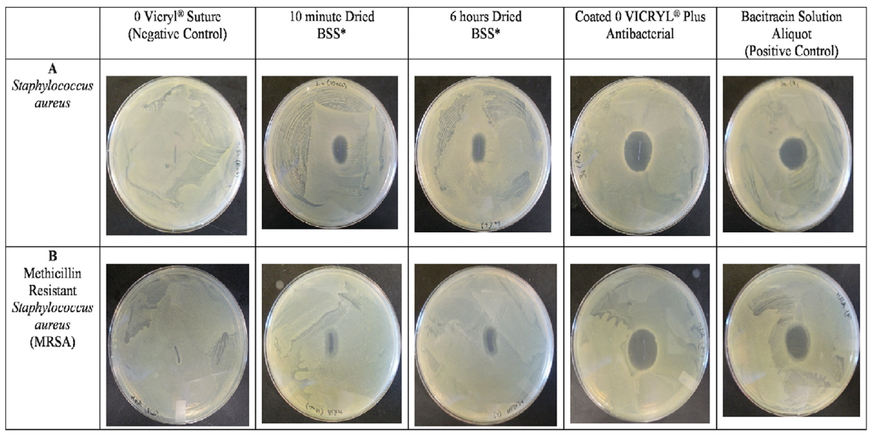
4.3.3. Application of Antibiotics in Designing Anti-Biofilm Surfaces

Antibiotics are a group of antibacterial agents, and can be incorporated into coatings to provide bactericidal surfaces. For instance, implant-related infections can be treated by implants that are coated with poly(D, L-Lactide) (PDLLA) loaded with gentamicin [226]. For example, Lucke et al. [227] proved that a gentamicin-loaded poly (d, l-lactide) (PDLLA) coating on orthopaedic devices could prevent implant-related osteomyelitis as an infection of the bone. In addition, Garvin et al. [228] confirmed that osteomyelitis infection was eradicated in tibiae of all mongrel dogs when they were treated with a polylactide/polyglycolide implant containing 100 milligrams of gentamicin. In another study, researchers prepared a vancomycin-loaded multilayer coating, consisting of montmorillonite and poly-L-lysine, to treat bone infections can kill more than 99.99% of S. aureus in 24 h [229].
The most common nosocomial infections are surgical site infections (SSI) which can be affected by suture materials. In this regard, researchers have studied the effect of triclosan coating (an antibacterial and antifungal agent) on two types of absorbable sutures, including Vicryl (manufactured by Ethicon Inc., Raritan, NJ, USA) and PDS® [210]. When coated with triclosan, both sutures could better reduce in vitro colonization of several strains of bacteria in comparison to control sutures [230].
In another study, the effect of triclosan coating was compared with bacitracin coating on antimicrobial activity of sutures [231]. In this study, the zone of inhibition around sutures is compared using Staphylococcus aureus and methicillin-resistant Staphylococcus aureus in different five groups, as illustrated in Figure 12. These groups include (1) sterile VICRYL® suture (without triclosan and Bacitracin) as the negative control, (2) Bacitracin-soaked suture (BSS) for 1 h and dried for 10 min, (3) BSS dried for 6 h, (4) VICRYL® Plus (triclosan-coated VICRYL), and (5) a Bacitracin solution as the positive control. Different drying times were considered to ensure that excess Bacitracin solution does not have any effect on measured antimicrobial activity. Although BSS sutures (dried for 10 min and 6 h) showed a significantly larger zone of inhibition compared to the negative control, they exhibited a smaller inhibition zone in comparison to the positive control or triclosan-coated VICRYL (VICRYL® Plus, currently on the market) and Bacitracin solution [231].
Hoshino et al. [232] used conventional polyglactin 910 sutures (VICRYL® suture) and triclosan-coated sutures (triclosan-coated VICRYL Plus) for the fascia closure in digestive tract surgery, and studied the effect of antibacterial coating on wound infection after surgery. Their results proved that triclosan-coated polyglactin sutures decrease surgical site infections in the study group. Wound infection developed in 6.6% of the patients receiving triclosan-coated VICRYL Plus, while 12.2% of patients that received sutures without triclosan-coating developed wound infection [232]. Zhang et al. [233] coated silver NPs on absorbable sutures by a layer-by-layer deposition method. They used the silver NPS-coated sutures for ideal anastomosis in mice. The silver NPS-coated suture showed in vitro antibacterial efficacy, significantly better collagen deposition in the anastomotic tissue, and less inflammatory cell infiltration (macrophage and neutrophil).
  • Triclosan-coated VICRYL Plus sutures

4.3.4. Application of Nanoparticles in Designing Anti-Biofilm Surfaces

Metallic NPs, including silver [234], gold [235], iron [236], titanium [237], zinc [238], copper [239], and their oxides, are also known as strong antibacterial agents. Their antibacterial effect is unlikely to produce antibacterial resistance. Hence, developing antibacterial NPs has become an effective method, since bacterial resistance is becoming a global health and development threat [240]. Rosenberg et al. [241] prepared surfaces coated with nano-zinc oxide (nano-ZnO) and nano-zinc oxide/silver (nano-ZnO/Ag) composites as nano-enabled surfaces, and showed that, in static oligotrophic conditions, colonization of E. coli and S. aureus was decreased.
To remove the pathogenic bacteria from groundwater sources, researchers designed filters by depositing silver NPs on different substrates including zeolite, sand, fibreglass, anion, and cation resin [242]. They proved that all filters were capable of decreasing the concentration of E. coli from synthetic water. Moreover, a Ag/cation resin substrate with complete (100%) removal of all targeted bacteria can be used as a cost-effective filter for the disinfection of groundwater and production of safe drinking water [242].
Applerot et al. (2012) developed an anti-biofilm surface coating with depositing zinc oxide (ZnO) [243] NPs on glass, by a one-step ultrasound irradiation process. Using electron spin resonance (ESR), they proved that the anti-biofilm activity of ZnO NP coating was mediated through the formation of ROS. They suggested that ZnO NPs can be utilized in producing anti-biofilm surfaces, all of which are prone to bacterial colonization in medical and industrial settings [243]. Many other studies have been performed to evaluate the antibacterial activity of metallic NPs embedded in a range of polymers in coatings [134,135,136,137,138]. Halloysite nanotubes have been used as a substrate for metalizing its surface with antimicrobial metal oxide nanoparticles [244,245,246,247,248].
Non-metallic NPs, including carbon-based, silica-based, and polymeric NPs, also exhibit a high antimicrobial activity [249,250] and can be used in designing anti-bacterial and anti-biofilm materials.

4.3.5. Designing Anti-Biofilm Bulk Material

Apart from designing an anti-biofouling surface, in some cases, antibacterial agents must be incorporated into the bulk material to prevent biofilm formation. For example, antibiotics and NPs can be applied in bone cement and wound dressing materials to prevent biofilm formation in patience.
For the treatment of osteomyelitis, to provide a sustained release of the antibiotic for adequate periods, hydrophilized poly (methyl methacrylate) (PMMA) bone cement containing a hydrophilic additive and vancomycin was prepared [251]. The results and findings of this study indicate that this antibiotic-eluting PMMA bone cement releases the antibiotic for up to 11 weeks and prohibits the growth of S. aureus over six weeks, as required to treat osteomyelitis [230]. Many other studies applied antibiotics, such as vancomycin, tobramycin, and gentamycin, into the bone cement to effectively treat chronic osteomyelitis-resistant cases [252]. However, Scott et al. [253] reported that gentamycin-loaded, compared to tobramycin-loaded cement, is less effective for treating Pseudomonas aeruginosa infection in septic joint arthroplasty.
Different types of wounds, such as burns, surgical wounds, and diabetic foot ulcers, can be infected, burdening the health care system. Topical antimicrobial agents will prevent wound infection, and have many advantages over systemic antibiotics for treating infection. Hence, drug-eluting wound dressings and adhesives possess many advantages over systemic antibiotics regarding wound healing. They provide a high concentration and sustained release of antimicrobial agents at the site only in the presence of infection, avoid systemic antibiotics, and reduce the risk of antibiotic resistance [254]. For example, biodegradable antibiotic-eluting wound dressings based on a polyglyconate mesh coated with a porous poly (dl-lactic-co-glycolic acid) matrix have been designed to protect the wound against infection and reduce the need for frequent dressing changing [255]. In addition, a ceftazidime antibiotic drug was incorporated into the novel tissue adhesives based on gelatine and alginate to design new bioadhesives for wound closing applications [256]. In another study, a polyurethane nanofibre-based nanocomposite containing silver NPS/zinc oxide (ZnAg) composite NPS for antibacterial wound dressing applications demonstrated 100% bactericidal properties against all the tested strains [257].
In 2020, researchers developed an anti-biofilm composite by application of silver/copper (Ag/Cu) NPs on the surface of graphene oxide (GO) nanosheets, as shown in Figure 13 [258]. The surface of the Ag/Cu/GO composite was capable of eradicating bacterial species applied in their study. The efficiency of this composite in removing the antibiotic-resistant bacteria through topical application of Ag/Cu/GO on a biofilm-infected skin wound of ICR mice has been evaluated. According to Figure 14, compared to the control group (PBS-treated group), wound closure was similar between the penicillin–streptomycin- (P/S) and Ag/Cu/GO-treated groups, and accelerated in comparison to the controlled group.
Many other antibiotic-eluting and NP-containing wound dressings have been developed, and show remarkable antibacterial activity and wound healing [259,260,261,262,263,264,265,266,267,268].
Controlling the release of antibiotics or NPs from the wound dressing substrate attracts considerable attention to designing an anti-infection environment for wound healing. In this regard, Shi et al. [269] designed a hydrogel network structure capable of the light-triggered release of a conjugating ciprofloxacin antibiotic through a photo-cleavable linker. Upon irradiation of the hydrogel material with low-intensity UV light, native ciprofloxacin was released, and its antimicrobial activity on S. aureus was demonstrated. Table 2 represents a comparative analysis of various drug-eluting or NP-embedded wound dressings, or antibacterial solutions and their potential uses in different types of wounds.

5. Nanoparticle–Bacteria Interactions

Releasing metal ions is the inhibitory mechanism of NPs against different bacteria. To understand the antibacterial effect of NPs, as shown in Figure 15, the reaction of silver NPs (representative of a metallic nanoparticle) with bacteria can be considered as an example. Silver NPs release Ag+ ions through an oxidative mechanism. Ag+ ions are stable and solid bactericides, and represent antibacterial activity against Gram-positive and Gram-negative bacteria [285,286]. NPs directly or indirectly damage bacteria, and their antibacterial effect stems from two main routes. First, they can physically and directly damage bacterial cell walls, membranes, or organelles and change their permeability by forming pores in the cell membrane. Second, toxic ions and reactive oxygen species (ROS) can be produced by NPs, which interact with the cell’s DNA, modulate signal transduction pathways, and arrest the cell cycle [287]. The first step of the mechanism of bacterial inhibition is NPs adhesion onto the surface of bacteria that depends on their size, shape, structure, and surface characteristics such as charge, composition, and wettability.
For instance, silver NPs of different sizes (7, 29, and 89 nm) were synthesized, and researchers proved that the antibacterial activity of the silver NPs decreases with an increase in the particle size [288]. To evaluate the role of the surface electrical charge of silver NPs on their antibacterial activity against Gram-positive and Gram-negative bacteria, different silver NPs providing three different electrical surface charges (positive, neutral, and negative) were synthesized. It was observed that positively charged silver NPs possess the highest bactericidal activity against tested Gram-positive and Gram-negative bacteria [289].
While neutrally charged particles had intermediate antibacterial activity, the negatively charged silver NPs had minor antibacterial activity [268]. In addition, spherical and rod-shaped silver NPs displayed the weakest biocidal action compared to truncated triangular silver NPs with a lattice plane [290]. However, NPs can have a genotoxicity or cytotoxicity effect on human cells. NPs’ mechanism of toxicity is similar to their antibacterial activity. For example, silver NPs can not only damage human cell surfaces and organelles, but can also generate ROS and cause genetic material damage [291]. NPs’ toxicity is directly controlled by their dose, concentration, exposure route, size, and frequency [291]. Hence, if they are going to be used for biomedical applications, it is essential to design NPs with optimum features that are toxic for bacteria but safe for human cells [292].

6. Future Directions

The association of the microbial biofilm community with pathogenic forms of human diseases and many life-threatening infections provides a compelling need for designing more effective antibacterial materials and surfaces. Systemic antibiotic therapy is not effective in infection prevention and treatment. Although significant research has been directed toward developing antibacterial substances and materials to prevent biofilm formation, cytotoxic effects of antibiotics on human cells and accelerating antibiotic resistance remain of concern.
There is a need to design advanced technologies for rapid and accurate detection of biofilm formation [293,294]. This includes exploring molecular-based methods, biosensors, and imaging technologies to enhance surveillance and early intervention capabilities.
Another future direction involves the implementation of innovative antimicrobial strategies, such as development of novel antimicrobial agents and antibiotics [295,296]. This may involve investigating antimicrobial peptides, surface modifications, and nanotechnology-based approaches to inhibit biofilm formation and enhance the effectiveness of disinfection techniques. For example, designing safe antibacterial NPs that are toxic for bacteria but safe for specific human cells will open new and unprecedented avenues for future research [297].
Future research efforts will focus on targeted biofilm prevention and control [298]. A deep understanding of biofilm formation mechanisms will drive the development of targeted interventions for biofilm prevention and disruption. This includes the design of antimicrobial surfaces, the use of biofilm-disrupting enzymes, and the exploration of microbial communication and signalling pathways for the development of specific therapies.
The future direction may involve the integration of comprehensive biofilm management strategies into industry practices across healthcare, food processing, industrial manufacturing, marine industries, and sanitation sectors [299]. This will entail the establishment of industry-wide guidelines, training programs, and standard operating procedures to minimize microbial contamination risks and improve sanitation practices.
Collaboration between academia, industry, and regulatory bodies will play a crucial role in addressing the challenges of biofilm-related issues. Sharing knowledge, best practices and research findings will facilitate the development of effective strategies, guidelines, and policies across different sectors.
It is worth mentioning that biofilm management techniques will be focused on developing environmentally friendly approaches in the future [300]. This includes exploring sustainable and non-toxic disinfection methods, bio-based materials, and eco-friendly biofilm control strategies that minimize the impact on human health and the environment.
By pursuing these future directions, we can enhance our understanding of microbial contamination and biofilm formation to control its negative impacts on healthcare, food safety, industrial processes, marine industries, and sanitation practices.
Nanoparticles, although showing practical effects on preventing and treating biofilms, are also toxic for human cells. The potential toxicity of NPs on human cells depends on the application site in the human body, cell type, and NPs’ physical and chemical characteristics such as size, dose, substance type, structure, and surface properties [272]. In addition, there also remains the potential harmful impact of nanoparticles on the environment.

7. Conclusions

Biofilms are self-organized and autonomously replicating microbial communities, having different bacterial colonies or single types of cells. Biofilm formation occurs in five main stages: attachment of cells to the surface, microcolony formation, proliferation, maturation, and dispersion. These complex microbiome structures are strongly protected by EPS as a self-produced and environmentally enriched matrix and show high antibiotic resistance. In addition, the EPS allows bacteria to communicate with each other through QS and autoinducer secretion to sense and control population density.
Biofilm formation is a persistent complication in medical and non-medical fields, and can cause many health or industrial problems. In non-medical industries, many environmental, industrial, and human health risks can also cause by biofilm formation. For example, the presence of biofilm in the water distribution system can decrease the water quality, release pipe material and contaminants into the water, increase the corrosion of pipe materials, and cause human health problems due to the distribution of pathogenic bacteria. In the food industry, they can increase the corrosion of the metallic surfaces in food manufacturing surfaces or generate unpleasant odours and tastes in products. In marine-based industries, biofilms can cause environmental and economic problems by increasing fuel consumption, CO2 emission, and deterioration of protective coatings.
Bacterial biofilms are responsible for 80% of chronic human body infections [273]. Healthcare-associated infections can be classified as non-device-related and device-related infections. Non-device-related infections can be caused by colonizing microorganisms on the teeth, skin, and mucosa [66]. For example, chronic surgical, diabetic, and burn wound infections are examples of non-device-related infections. In contrast, device-related infections are caused by biofilm formation on temporary or permanent implantable medical devices.
The formation of conditioning films is the prerequisite step for biofilm formation, and it includes the adsorption of biomolecules from the surrounding environment to prepare the surface for bacterial adhesion. These macromolecules can be absorbed from human biological fluid, or sea or drinking water. The first step of biofilm formation is the attachment of bacteria to a favourable surface. After a successful attachment, cells start to multiply and produce biofilm matrix components. Hence, the first step is the most critical stage, and its prevention will inhibit bacterial proliferation and biofilm formation. As initial attachment is critical for forming a bacterial biofilm, there is a need to develop strategies to prevent or diminish biofilm formation.
We categorized the methods to prevent biofilm generation into two groups: designing anti-biofilm surfaces and designing anti-biofilm bulk materials. We believe that there are two main strategies to design anti-biofilm surfaces inspired by nature, and they include (1) designing cell-repellent surfaces and (2) bactericidal substrates. Other strategies can be counted as the subgroups of these two strategies. Protein- and cell-repellent surfaces prevent cell attachment, while bactericidal surfaces deactivate or kill the attached bacteria.
To prepare cell-repellent surfaces, surface physical and chemical characteristics, such as structure, roughness, charge, and chemical composition, are determinant factors of cell-surface interaction. Hence, unfavourable surface topography and chemistry will prevent the attachment of bacteria to the surface and, consequently, its proliferation. To develop bactericidal substrates, antibacterial agents, including antibiotics (such as antibiotics vancomycin, tobramycin, and gentamycin) and NPs (such as gold and silver NPs), can be applied on the surface through films or coatings.
Applying such antibacterial agents into the bulk material is also counted as an effective method in engineering anti-biofilm material for various applications. Drug-eluting bone cement wound dressings and sutures are examples of this category’s successful anti-biofilm and infection properties.

Author Contributions

Conceptualization, G.S., M.M., M.J.P.B., D.K.M., Original draft preparation, G.S.: Review and editing, G.S., M.M., M.J.P.B., D.K.M.; Supervision, D.K.M.; Project administration, D.K.M.; Funding acquisition, D.K.M. All authors have read and agreed to the published version of the manuscript.

Funding

This manuscript was funded by a NASA EPSCoR Rapid Response Research (R3) program award 22-2022 R30015.

Data Availability Statement

Not applicable.

Conflicts of Interest

The authors declare no conflict of interest.

References

  1. Nazir, R.; Zaffar, M.R.; Amin, I. Bacterial biofilms: The remarkable heterogeneous biological communities and nitrogen fixing microorganisms in lakes. Freshw. Microbiol. 2019, 307–340. [Google Scholar]
  2. Costa, O.Y.; Raaijmakers, J.M.; Kuramae, E.E. Microbial extracellular polymeric substances: Ecological function and impact on soil aggregation. Front. Microbiol. 2018, 9, 1636. [Google Scholar] [CrossRef] [PubMed] [Green Version]
  3. Chen, M.; Yu, Q.; Sun, H. Novel strategies for the prevention and treatment of biofilm related infections. Int. J. Mol. Sci. 2013, 14, 18488–18501. [Google Scholar] [CrossRef] [PubMed] [Green Version]
  4. Khatoon, Z.; McTiernan, C.D.; Suuronen, E.J.; Mah, T.-F.; Alarcon, E.I. Bacterial biofilm formation on implantable devices and approaches to its treatment and prevention. Heliyon 2018, 4, e01067. [Google Scholar] [CrossRef] [Green Version]
  5. Davies, D. Understanding biofilm resistance to antibacterial agents. Nat. Rev. Drug Discov. 2003, 2, 114–122. [Google Scholar] [CrossRef]
  6. Noguera, D.R.; Okabe, S.; Picioreanu, C. Biofilm modeling: Present status and future directions. Water Sci. Technol. 1999, 39, 273–278. [Google Scholar] [CrossRef]
  7. Swanson, T.; Wolcott, R.; Wallis, H.; Woodmansey, E. Understanding biofilm in practice: A global survey of health professionals. J. Wound Care 2017, 26, 426–440. [Google Scholar] [CrossRef] [PubMed]
  8. Di Martino, P. Extracellular polymeric substances, a key element in understanding biofilm phenotype. AIMS Microbiol. 2018, 4, 274. [Google Scholar] [CrossRef] [PubMed]
  9. Hall, M.R.; McGillicuddy, E.; Kaplan, L.J. Biofilm: Basic principles, pathophysiology, and implications for clinicians. Surg. Infect. 2014, 15, 1–7. [Google Scholar] [CrossRef]
  10. Srivastava, S.; Bhargava, A. Biofilms and human health. Biotechnol. Lett. 2016, 38, 1–22. [Google Scholar] [CrossRef]
  11. Büttner, H.; Mack, D.; Rohde, H. Structural basis of Staphylococcus epidermidis biofilm formation: Mechanisms and molecular interactions. Front. Cell. Infect. Microbiol. 2015, 5, 14. [Google Scholar]
  12. Irie, Y.; Borlee, B.R.; O’Connor, J.R.; Hill, P.J.; Harwood, C.S.; Wozniak, D.J.; Parsek, M.R. Self-produced exopolysaccharide is a signal that stimulates biofilm formation in Pseudomonas aeruginosa. Proc. Natl. Acad. Sci. USA 2012, 109, 20632–20636. [Google Scholar] [CrossRef] [Green Version]
  13. Rimondini, L.; Cochis, A.; Varoni, E.; Azzimonti, B.; Carrassi, A. Biofilm formation on implants and prosthetic dental materials. In Handbook of Bioceramics and Biocomposites; Elsevier: Amsterdam, The Netherlands, 2015; pp. 1–37. [Google Scholar]
  14. Bjarnsholt, T. The role of bacterial biofilms in chronic infections. Apmis 2013, 121, 1–58. [Google Scholar] [CrossRef] [PubMed]
  15. Del Pozo, J.; Patel, R. The challenge of treating biofilm-associated bacterial infections. Clin. Pharmacol. Ther. 2007, 82, 204–209. [Google Scholar] [CrossRef]
  16. VanEpps, J.S.; Younger, J.G. Implantable device related infection. Shock 2016, 46, 597. [Google Scholar] [CrossRef] [Green Version]
  17. De la Fuente-Núñez, C.; Reffuveille, F.; Fernández, L.; Hancock, R.E. Bacterial biofilm development as a multicellular adaptation: Antibiotic resistance and new therapeutic strategies. Curr. Opin. Microbiol. 2013, 16, 580–589. [Google Scholar] [CrossRef]
  18. Zheng, S.; Bawazir, M.; Dhall, A.; Kim, H.-E.; He, L.; Heo, J.; Hwang, G. Implication of surface properties, bacterial motility, and hydrodynamic conditions on bacterial surface sensing and their initial adhesion. Front. Bioeng. Biotechnol. 2021, 9, 643722. [Google Scholar] [CrossRef] [PubMed]
  19. Rummel, C.D.; Lechtenfeld, O.J.; Kallies, R.; Benke, A.; Herzsprung, P.; Rynek, R.; Wagner, S.; Potthoff, A.; Jahnke, A.; Schmitt-Jansen, M. Conditioning film and early biofilm succession on plastic surfaces. Environ. Sci. Technol. 2021, 55, 11006–11018. [Google Scholar] [CrossRef]
  20. Lorite, G.S.; Rodrigues, C.M.; De Souza, A.A.; Kranz, C.; Mizaikoff, B.; Cotta, M.A. The role of conditioning film formation and surface chemical changes on Xylella fastidiosa adhesion and biofilm evolution. J. Colloid Interface Sci. 2011, 359, 289–295. [Google Scholar] [CrossRef] [PubMed] [Green Version]
  21. Hwang, G.; Liang, J.; Kang, S.; Tong, M.; Liu, Y. The role of conditioning film formation in Pseudomonas aeruginosa PAO1 adhesion to inert surfaces in aquatic environments. Biochem. Eng. J. 2013, 76, 90–98. [Google Scholar] [CrossRef]
  22. Bhagwat, G.; O’Connor, W.; Grainge, I.; Palanisami, T. Understanding the fundamental basis for biofilm formation on plastic surfaces: Role of conditioning films. Front. Microbiol. 2021, 12, 687118. [Google Scholar] [CrossRef]
  23. Talluri, S.N.; Winter, R.M.; Salem, D.R. Conditioning film formation and its influence on the initial adhesion and biofilm formation by a cyanobacterium on photobioreactor materials. Biofouling 2020, 36, 183–199. [Google Scholar] [CrossRef]
  24. Cooksey, K.; Wigglesworth-Cooksey, B. Adhesion of bacteria and diatoms to surfaces in the sea: A review. Aquat. Microb. Ecol. 1995, 9, 87–96. [Google Scholar] [CrossRef]
  25. Ammar, Y.; Swailes, D.; Bridgens, B.; Chen, J. Influence of surface roughness on the initial formation of biofilm. Surf. Coat. Technol. 2015, 284, 410–416. [Google Scholar] [CrossRef] [Green Version]
  26. Spengler, C.; Nolle, F.; Mischo, J.; Faidt, T.; Grandthyll, S.; Thewes, N.; Koch, M.; Müller, F.; Bischoff, M.; Klatt, M.A. Strength of bacterial adhesion on nanostructured surfaces quantified by substrate morphometry. Nanoscale 2019, 11, 19713–19722. [Google Scholar] [CrossRef] [Green Version]
  27. Cao, L.; Chen, X.; Peng, Y. The adsorption and orientation of frother surfactants on heterogeneous wetting surfaces. Appl. Surf. Sci. 2021, 548, 149225. [Google Scholar] [CrossRef]
  28. Krasowska, A.; Sigler, K. How microorganisms use hydrophobicity and what does this mean for human needs? Front. Cell. Infect. Microbiol. 2014, 4, 112. [Google Scholar] [CrossRef] [PubMed] [Green Version]
  29. Riau, A.K.; Aung, T.T.; Setiawan, M.; Yang, L.; Yam, H.F.G.; Beuerman, W.R.; Venkatraman, S.S.; Mehta, S.J. Surface immobilization of nano-silver on polymeric medical devices to prevent bacterial biofilm formation. Pathogens 2019, 8, 93. [Google Scholar] [CrossRef] [PubMed] [Green Version]
  30. Aiyer, K.S.; Vijayakumar, B.; Vishwanathan, A. The enigma of biofilms. Curr. Sci. 2018, 115, 204–205. [Google Scholar] [CrossRef]
  31. Wang, L.; Fan, D.; Chen, W.; Terentjev, E.M. Bacterial growth, detachment and cell size control on polyethylene terephthalate surfaces. Sci. Rep. 2015, 5, 15159. [Google Scholar] [CrossRef] [Green Version]
  32. Lagage, V.; Uphoff, S. Filming flagella and pili in action. Nat. Rev. Microbiol. 2018, 16, 584. [Google Scholar] [CrossRef] [PubMed]
  33. Mérian, T.; Goddard, J.M. Advances in nonfouling materials: Perspectives for the food industry. J. Agric. Food Chem. 2012, 60, 2943–2957. [Google Scholar] [CrossRef] [PubMed]
  34. Feng, G.; Cheng, Y.; Wang, S.-Y.; Borca-Tasciuc, D.A.; Worobo, R.W.; Moraru, C.I. Bacterial attachment and biofilm formation on surfaces are reduced by small-diameter nanoscale pores: How small is small enough? npj Biofilms Microbiomes 2015, 1, 1–9. [Google Scholar] [CrossRef] [PubMed] [Green Version]
  35. Abdallah, M.; Benoliel, C.; Drider, D.; Dhulster, P.; Chihib, N.-E. Biofilm formation and persistence on abiotic surfaces in the context of food and medical environments. Arch. Microbiol. 2014, 196, 453–472. [Google Scholar] [CrossRef]
  36. Greer, H.M.; Overton, K.; Ferguson, M.A.; Spain, E.M.; Darling, L.E.; Núñez, M.E.; Volle, C.B. Extracellular Polymeric Substance Protects Some Cells in an Escherichia coli Biofilm from the Biomechanical Consequences of Treatment with Magainin 2. Microorganisms 2021, 9, 976. [Google Scholar] [CrossRef]
  37. Mukherjee, S.; Bassler, B.L. Bacterial quorum sensing in complex and dynamically changing environments. Nat. Rev. Microbiol. 2019, 17, 371–382. [Google Scholar] [CrossRef] [PubMed]
  38. Schaechter, M. Encyclopedia of Microbiology; Academic Press: Cambridge, MA, USA, 2009. [Google Scholar]
  39. Strycharz-Glaven, S.M.; Snider, R.M.; Guiseppi-Elie, A.; Tender, L.M. On the electrical conductivity of microbial nanowires and biofilms. Energy Environ. Sci. 2011, 4, 4366–4379. [Google Scholar] [CrossRef]
  40. Neu, J.; Shipps, C.C.; Guberman-Pfeffer, M.J.; Shen, C.; Srikanth, V.; Spies, J.A.; Kirchhofer, N.D.; Yalcin, S.E.; Brudvig, G.W.; Batista, V.S. Microbial biofilms as living photoconductors due to ultrafast electron transfer in cytochrome OmcS nanowires. Nat. Commun. 2022, 13, 5150. [Google Scholar] [CrossRef]
  41. Reguera, G. Microbial nanowires and electroactive biofilms. FEMS Microbiol. Ecol. 2018, 94, fiy086. [Google Scholar] [CrossRef] [PubMed]
  42. Ye, Y.; Liu, X.; Nealson, K.H.; Rensing, C.; Qin, S.; Zhou, S. Dissecting the structural and conductive functions of nanowires in Geobacter sulfurreducens electroactive biofilms. mBio 2022, 13, e03822-21. [Google Scholar] [CrossRef] [PubMed]
  43. Sauer, K.; Cullen, M.; Rickard, A.; Zeef, L.; Davies, D.G.; Gilbert, P. Characterization of nutrient-induced dispersion in Pseudomonas aeruginosa PAO1 biofilm. J. Bacteriol. 2004, 186, 7312–7326. [Google Scholar] [CrossRef] [Green Version]
  44. Machineni, L.; Rajapantul, A.; Nandamuri, V.; Pawar, P.D. Influence of nutrient availability and quorum sensing on the formation of metabolically inactive microcolonies within structurally heterogeneous bacterial biofilms: An individual-based 3D cellular automata model. Bull. Math. Biol. 2017, 79, 594–618. [Google Scholar] [CrossRef]
  45. Landini, P.; Antoniani, D.; Burgess, J.G.; Nijland, R. Molecular mechanisms of compounds affecting bacterial biofilm formation and dispersal. Appl. Microbiol. Biotechnol. 2010, 86, 813–823. [Google Scholar] [CrossRef] [PubMed]
  46. Vilain, S.; Cosette, P.; Zimmerlin, I.; Dupont, J.-P.; Junter, G.-A.; Jouenne, T. Biofilm proteome: Homogeneity or versatility? J. Proteome Res. 2004, 3, 132–136. [Google Scholar] [CrossRef] [PubMed]
  47. Bowler, P.; Murphy, C.; Wolcott, R. Biofilm exacerbates antibiotic resistance: Is this a current oversight in antimicrobial stewardship? Antimicrob. Resist. Infect. Control 2020, 9, 1–5. [Google Scholar] [CrossRef] [PubMed]
  48. Yi, L.; Jin, M.; Li, J.; Grenier, D.; Wang, Y. Antibiotic resistance related to biofilm formation in Streptococcus suis. Appl. Microbiol. Biotechnol. 2020, 104, 8649–8660. [Google Scholar] [CrossRef] [PubMed]
  49. Fauzia, K.A.; Miftahussurur, M.; Syam, A.F.; Waskito, L.A.; Doohan, D.; Rezkitha, Y.A.A.; Matsumoto, T.; Tuan, V.P.; Akada, J.; Yonezawa, H. Biofilm formation and antibiotic resistance phenotype of Helicobacter pylori clinical isolates. Toxins 2020, 12, 473. [Google Scholar] [CrossRef]
  50. Pereira, R.; dos Santos Fontenelle, R.; De Brito, E.; De Morais, S. Biofilm of Candida albicans: Formation, regulation and resistance. J. Appl. Microbiol. 2021, 131, 11–22. [Google Scholar] [CrossRef]
  51. Abriat, C.; Gazil, O.; Heuzey, M.-C.; Daigle, F.; Virgilio, N. The polymeric matrix composition of Vibrio cholerae biofilms modulate resistance to silver nanoparticles prepared by hydrothermal synthesis. ACS Appl. Mater. Interfaces 2021, 13, 35356–35364. [Google Scholar] [CrossRef]
  52. Uruén, C.; Chopo-Escuin, G.; Tommassen, J.; Mainar-Jaime, R.C.; Arenas, J. Biofilms as promoters of bacterial antibiotic resistance and tolerance. Antibiotics 2020, 10, 3. [Google Scholar] [CrossRef]
  53. Goel, N.; Fatima, S.W.; Kumar, S.; Sinha, R.; Khare, S.K. Antimicrobial resistance in biofilms: Exploring marine actinobacteria as a potential source of antibiotics and biofilm inhibitors. Biotechnol. Rep. 2021, 30, e00613. [Google Scholar] [CrossRef]
  54. Piras, C.; Di Ciccio, P.A.; Soggiu, A.; Greco, V.; Tilocca, B.; Costanzo, N.; Ceniti, C.; Urbani, A.; Bonizzi, L.; Ianieri, A.S. aureus biofilm protein expression linked to antimicrobial resistance: A proteomic study. Animals 2021, 11, 966. [Google Scholar] [CrossRef] [PubMed]
  55. Gilbert, P.; Allison, D.; McBain, A. Biofilms in vitro and in vivo: Do singular mechanisms imply cross-resistance? J. Appl. Microbiol. 2002, 92, 98S–110S. [Google Scholar] [CrossRef]
  56. Yadav, M.K.; Song, J.-J.; Singh, B.P.; Vidal, J.E. Microbial biofilms and human disease: A concise review. New Future Dev. Microb. Biotechnol. Bioeng. Microb. Biofilms 2020, 1, 1–13. [Google Scholar]
  57. Li, Y.; Xiao, P.; Wang, Y.; Hao, Y. Mechanisms and control measures of mature biofilm resistance to antimicrobial agents in the clinical context. ACS Omega 2020, 5, 22684–22690. [Google Scholar] [CrossRef] [PubMed]
  58. Donlan, R.M. Biofilm formation: A clinically relevant microbiological process. Clin. Infect. Dis. 2001, 33, 1387–1392. [Google Scholar] [CrossRef] [PubMed] [Green Version]
  59. Kostakioti, M.; Hadjifrangiskou, M.; Hultgren, S.J. Bacterial biofilms: Development, dispersal, and therapeutic strategies in the dawn of the postantibiotic era. Cold Spring Harb. Perspect. Med. 2013, 3, a010306. [Google Scholar] [CrossRef] [PubMed] [Green Version]
  60. Davey, M.E.; O’toole, G.A. Microbial biofilms: From ecology to molecular genetics. Microbiol. Mol. Biol. Rev. 2000, 64, 847–867. [Google Scholar] [CrossRef] [Green Version]
  61. Swartjes, J.J.; Das, T.; Sharifi, S.; Subbiahdoss, G.; Sharma, P.K.; Krom, B.P.; Busscher, H.J.; van der Mei, H.C. A functional DNase I coating to prevent adhesion of bacteria and the formation of biofilm. Adv. Funct. Mater. 2013, 23, 2843–2849. [Google Scholar] [CrossRef]
  62. Costerton, J.W.; Lewandowski, Z.; Caldwell, D.E.; Korber, D.R.; Lappin-Scott, H.M. Microbial biofilms. Annu. Rev. Microbiol. 1995, 49, 711–745. [Google Scholar] [CrossRef]
  63. Stewart, P.S. Diffusion in biofilms. J. Bacteriol. 2003, 185, 1485–1491. [Google Scholar] [CrossRef] [PubMed] [Green Version]
  64. Smirnova, T.; Didenko, L.; Azizbekyan, R.; Romanova, Y.M. Structural and functional characteristics of bacterial biofilms. Microbiology 2010, 79, 413–423. [Google Scholar] [CrossRef]
  65. Saini, R.; Giri, P.; Saini, S.; Saini, S. Dental Plaque: A Complex Biofilm. Pravara Med. Rev. 2015, 7, 9–14. [Google Scholar]
  66. Carette, J.; Nachtergael, A.; Duez, P.; El Jaziri, M.; Rasamiravaka, T. Natural compounds inhibiting Pseudomonas aeruginosa biofilm formation by targeting quorum sensing circuitry. Bact. Biofilms 2020. [Google Scholar] [CrossRef] [Green Version]
  67. Fanning, S.; Mitchell, A.P. Fungal biofilms. PLoS Pathog. 2012, 8, e1002585. [Google Scholar] [CrossRef] [Green Version]
  68. Ruiz, A.; Herráez, M.; Costa-Gutierrez, S.B.; Molina-Henares, M.A.; Martínez, M.J.; Espinosa-Urgel, M.; Barriuso, J. The architecture of a mixed fungal–bacterial biofilm is modulated by quorum-sensing signals. Environ. Microbiol. 2021, 23, 2433–2447. [Google Scholar] [CrossRef]
  69. Gutiérrez, D.; Rodríguez-Rubio, L.; Martínez, B.; Rodríguez, A.; García, P. Bacteriophages as weapons against bacterial biofilms in the food industry. Front. Microbiol. 2016, 7, 825. [Google Scholar] [CrossRef] [Green Version]
  70. Colavecchio, A.; Cadieux, B.; Lo, A.; Goodridge, L.D. Bacteriophages contribute to the spread of antibiotic resistance genes among foodborne pathogens of the Enterobacteriaceae family—A review. Front. Microbiol. 2017, 8, 1108. [Google Scholar] [CrossRef] [Green Version]
  71. Khelissa, S.O.; Abdallah, M.; Jama, C.; Faille, C.; Chihib, N.-E. Bacterial contamination and biofilm formation on abiotic surfaces and strategies to overcome their persistence. J. Mater. Environ. Sci. 2017, 8, 3326–3346. [Google Scholar]
  72. Almaguer-Flores, A. Biofilms in the oral environment. In Bio-Tribocorrosion in Biomaterials and Medical Implants; Elsevier: Amsterdam, The Netherlands, 2013; pp. 169–186. [Google Scholar]
  73. Archibald, L.; Phillips, L.; Monnet, D.; McGowan, J.E., Jr.; Tenover, F.; Gaynes, R. Antimicrobial resistance in isolates from inpatients and outpatients in the United States: Increasing importance of the intensive care unit. Clin. Infect. Dis. 1997, 24, 211–215. [Google Scholar] [CrossRef] [Green Version]
  74. Berbari, E.F.; Hanssen, A.D.; Duffy, M.C.; Steckelberg, J.M.; Ilstrup, D.M.; Harmsen, W.S.; Osmon, D.R. Risk factors for prosthetic joint infection: Case-control study. Clin. Infect. Dis. 1998, 27, 1247–1254. [Google Scholar] [CrossRef]
  75. Almqvist, M.; Mattsson, G.; Razmi, R.; Magnusson, P. Cardiac Implantable Electronic Device-Related Infections. In Infective Endocarditis; IntechOpen: London, UK, 2019. [Google Scholar]
  76. Kranz, J.; Schmidt, S.; Wagenlehner, F.; Schneidewind, L. Catheter-associated urinary tract infections in adult patients: Preventive strategies and treatment options. Dtsch. Ärzteblatt Int. 2020, 117, 83. [Google Scholar]
  77. Papazian, L.; Klompas, M.; Luyt, C.-E. Ventilator-associated pneumonia in adults: A narrative review. Intensive Care Med. 2020, 46, 888–906. [Google Scholar] [CrossRef] [Green Version]
  78. Jacqz-Aigrain, E.; Choonara, I. Paediatric Clinical Pharmacology; CRC Press: Boca Raton, FL, USA, 2021. [Google Scholar]
  79. Kramer, G.; Klingler, H.C.; Steiner, G.E. Role of bacteria in the development of kidney stones. Curr. Opin. Urol. 2000, 10, 35–38. [Google Scholar] [CrossRef] [PubMed]
  80. Saini, R.; Marawar, P.; Shete, S.; Saini, S. Periodontitis, a true infection. J. Glob. Infect. Dis. 2009, 1, 149. [Google Scholar] [CrossRef] [PubMed]
  81. Rajani, R.; Klein, J.L. Infective endocarditis: A contemporary update. Clin. Med. 2020, 20, 31. [Google Scholar] [CrossRef]
  82. Saiman, L.; Siegel, J. Infection control in cystic fibrosis. Clin. Microbiol. Rev. 2004, 17, 57–71. [Google Scholar] [CrossRef] [Green Version]
  83. Mancl, K.A.; Kirsner, R.S.; Ajdic, D. Wound biofilms: Lessons learned from oral biofilms. Wound Repair Regen. 2013, 21, 352–362. [Google Scholar] [CrossRef] [Green Version]
  84. Brandwein, M.; Steinberg, D.; Meshner, S. Microbial biofilms and the human skin microbiome. NPJ Biofilms Microbiomes 2016, 2, 3. [Google Scholar] [CrossRef] [Green Version]
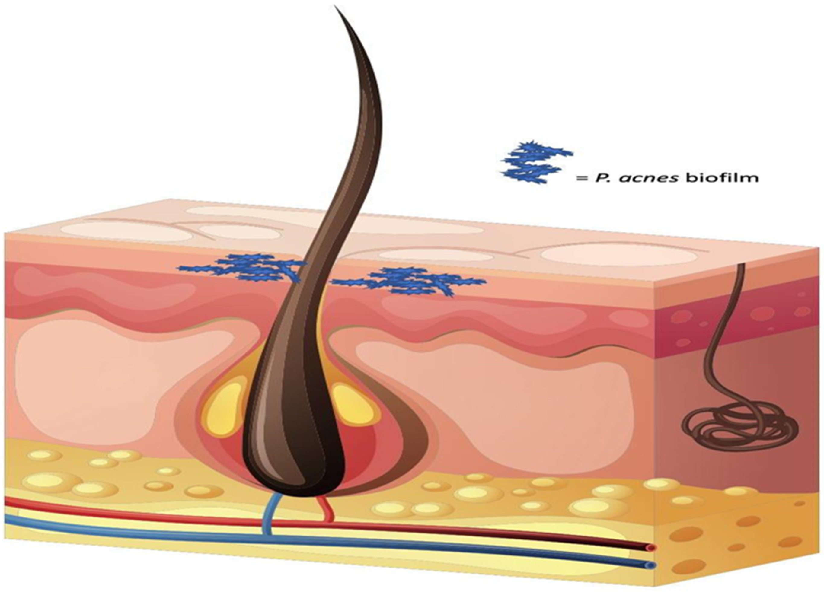
  85. Coenye, T.; Honraet, K.; Rossel, B.; Nelis, H.J. Biofilms in skin infections: Propionibacterium acnes and acne vulgaris. Infect. Disord. -Drug Targets Former. Curr. Drug Targets-Infect. Disord. 2008, 8, 156–159. [Google Scholar] [CrossRef]
  86. Di Domenico, E.; Cavallo, I.; Bordignon, V.; Prignano, G.; Sperduti, I.; Gurtner, A.; Trento, E.; Toma, L.; Pimpinelli, F.; Capitanio, B. Inflammatory cytokines and biofilm production sustain Staphylococcus aureus outgrowth and persistence: A pivotal interplay in the pathogenesis of Atopic Dermatitis. Sci. Rep. 2018, 8, 9573. [Google Scholar] [CrossRef] [PubMed] [Green Version]
  87. Rajpaul, K. Biofilm in wound care. Br. J. Community Nurs. 2015, 20, S6–S11. [Google Scholar] [CrossRef] [PubMed]
  88. Shineh, G.; Kordestani, S.S.; Tahriri, M.; Tayebi, L. Evaluation of L929 cell morphology on anthocyanin-containing gelatin-based hydrogel for early detection of infection. Bio-Des. Manuf. 2019, 2, 181–186. [Google Scholar] [CrossRef]
  89. Agyingi, E.; Maggelakis, S.; Ross, D. The effect of bacteria on epidermal wound healing. Math. Model. Nat. Phenom. 2010, 5, 28–39. [Google Scholar] [CrossRef] [Green Version]
  90. Rossi, E.; La Rosa, R.; Bartell, J.A.; Marvig, R.L.; Haagensen, J.A.; Sommer, L.M.; Molin, S.; Johansen, H.K. Pseudomonas aeruginosa adaptation and evolution in patients with cystic fibrosis. Nat. Rev. Microbiol. 2021, 19, 331–342. [Google Scholar] [CrossRef]
  91. Costerton, J.W.; Stewart, P.S.; Greenberg, E.P. Bacterial biofilms: A common cause of persistent infections. Science 1999, 284, 1318–1322. [Google Scholar] [CrossRef] [Green Version]
  92. Roberts, C.G. The role of biofilms in reprocessing medical devices. Am. J. Infect. Control 2013, 41, S77–S80. [Google Scholar] [CrossRef]
  93. Pearle, M.S.; Roehrborn, C.G. Antimicrobial prophylaxis prior to shock wave lithotripsy in patients with sterile urine before treatment: A meta-analysis and cost-effectiveness analysis. Urology 1997, 49, 679–686. [Google Scholar] [CrossRef]
  94. Ciofu, O.; Rojo-Molinero, E.; Macià, M.D.; Oliver, A. Antibiotic treatment of biofilm infections. Apmis 2017, 125, 304–319. [Google Scholar] [CrossRef] [Green Version]
  95. Lee, S.W.; Phillips, K.S.; Gu, H.; Kazemzadeh-Narbat, M.; Ren, D. How microbes read the map: Effects of implant topography on bacterial adhesion and biofilm formation. Biomaterials 2021, 268, 120595. [Google Scholar] [CrossRef]
  96. Pipattanachat, S.; Qin, J.; Rokaya, D.; Thanyasrisung, P.; Srimaneepong, V. Biofilm inhibition and bactericidal activity of NiTi alloy coated with graphene oxide/silver nanoparticles via electrophoretic deposition. Sci. Rep. 2021, 11, 14008. [Google Scholar] [CrossRef] [PubMed]
  97. Piktel, E.; Suprewicz, Ł.; Depciuch, J.; Chmielewska, S.; Skłodowski, K.; Daniluk, T.; Król, G.; Kołat-Brodecka, P.; Bijak, P.; Pajor-Świerzy, A. Varied-shaped gold nanoparticles with nanogram killing efficiency as potential antimicrobial surface coatings for the medical devices. Sci. Rep. 2021, 11, 12546. [Google Scholar] [CrossRef] [PubMed]
  98. Tang, M.; Chen, C.; Zhu, J.; Allcock, H.R.; Siedlecki, C.A.; Xu, L.-C. Inhibition of bacterial adhesion and biofilm formation by a textured fluorinated alkoxyphosphazene surface. Bioact. Mater. 2021, 6, 447–459. [Google Scholar] [CrossRef] [PubMed]
  99. Nagay, B.E.; Dini, C.; Cordeiro, J.M.; Ricomini-Filho, A.n.P.; De Avila, E.D.; Rangel, E.C.; Da Cruz, N.C.; Barão, V.A. Visible-light-induced photocatalytic and antibacterial activity of tio2 codoped with nitrogen and bismuth: New perspectives to control implant-biofilm-related diseases. ACS Appl. Mater. Interfaces 2019, 11, 18186–18202. [Google Scholar] [CrossRef]
  100. Servais, P.; Laurent, P.; Randon, G. Comparison of the bacterial dynamics in various French distribution systems. Aqua-Lond. Oxf. -J. Int. Water Supply Assoc. 1995, 44, 10. [Google Scholar]
  101. September, S.; Els, F.; Venter, S.; Brözel, V. Prevalence of bacterial pathogens in biofilms of drinking water distribution systems. J. Water Health 2007, 5, 219–227. [Google Scholar] [CrossRef] [Green Version]
  102. Lee, S.H.; O’Connor, J.T.; Banerji, S.K. Biologically mediated corrosion and its effects on water quality in distribution systems. J. Am. Water Work. Assoc. 1980, 72, 636–645. [Google Scholar] [CrossRef]
  103. Levy, R.V.; Hart, F.L.; Cheetham, R.D. Occurrence and public health significance of invertebrates in drinking water systems. J. Am. Water Work. Assoc. 1986, 78, 105–110. [Google Scholar] [CrossRef]
  104. Tsvetanova, Z. Study of biofilm formation on different pipe materials in a model of drinking water distribution system and its impact on microbiological water quality. In Chemicals as Intentional and Accidental Global Environmental Threats; Springer: Dordrecht, The Netherlands, 2006; pp. 463–468. [Google Scholar]
  105. Hallam, N.; West, J.; Forster, C.; Simms, J. The potential for biofilm growth in water distribution systems. Water Res. 2001, 35, 4063–4071. [Google Scholar] [CrossRef]
  106. Ren, H.; Wang, W.; Liu, Y.; Liu, S.; Lou, L.; Cheng, D.; He, X.; Zhou, X.; Qiu, S.; Fu, L. Pyrosequencing analysis of bacterial communities in biofilms from different pipe materials in a city drinking water distribution system of East China. Appl. Microbiol. Biotechnol. 2015, 99, 10713–10724. [Google Scholar] [CrossRef]
  107. Cowle, M.W.; Webster, G.; Babatunde, A.O.; Bockelmann-Evans, B.N.; Weightman, A.J. Impact of flow hydrodynamics and pipe material properties on biofilm development within drinking water systems. Environ. Technol. 2020, 41, 3732–3744. [Google Scholar] [CrossRef] [PubMed] [Green Version]
  108. Tsagkari, E.; Sloan, W. Turbulence accelerates the growth of drinking water biofilms. Bioprocess Biosyst. Eng. 2018, 41, 757–770. [Google Scholar] [CrossRef] [Green Version]
  109. Ercumen, A.; Gruber, J.S.; Colford, J.M., Jr. Water distribution system deficiencies and gastrointestinal illness: A systematic review and meta-analysis. Environ. Health Perspect. 2014, 122, 651–660. [Google Scholar] [CrossRef] [PubMed] [Green Version]
  110. Semenza, J.C.; Roberts, L.; Henderson, A.; Bogan, J.; Rubin, C. Water distribution system and diarrheal disease transmission: A case study in Uzbekistan. Am. J. Trop. Med. Hyg. 1998, 59, 941–946. [Google Scholar] [CrossRef] [PubMed]
  111. Percival, S.L.; Chalmers, R.; Hunter, P.R.; Sellwood, J.; Wyn-Jones, P. Microbiology of Waterborne Diseases; Elsevier: Amsterdam, The Netherlands; Academic Press: Cambridge, MA, USA, 2004; Volume 92101. [Google Scholar]
  112. Payment, P. Epidemiology of endemic gastrointestinal and respiratory diseases: Incidence, fraction attributable to tap water and costs to society. Water Sci. Technol. 1997, 35, 7–10. [Google Scholar] [CrossRef]
  113. Simoes, L.C.; Simões, M. Biofilms in drinking water: Problems and solutions. Rsc Adv. 2013, 3, 2520–2533. [Google Scholar] [CrossRef] [Green Version]
  114. de Faria, A.F.; Martinez, D.S.T.; Meira, S.M.M.; de Moraes, A.C.M.; Brandelli, A.; Souza Filho, A.G.; Alves, O.L. Anti-adhesion and antibacterial activity of silver nanoparticles supported on graphene oxide sheets. Colloids Surf. B Biointerfaces 2014, 113, 115–124. [Google Scholar] [CrossRef]
  115. Shi, X.; Zhu, X. Biofilm formation and food safety in food industries. Trends Food Sci. Technol. 2009, 20, 407–413. [Google Scholar] [CrossRef]
  116. Wang, H.; Wang, H.; Xing, T.; Wu, N.; Xu, X.; Zhou, G. Removal of Salmonella biofilm formed under meat processing environment by surfactant in combination with bio-enzyme. LWT-Food Sci. Technol. 2016, 66, 298–304. [Google Scholar] [CrossRef]
  117. Mafu, A.A.; Plumety, C.; Deschênes, L.; Goulet, J. Adhesion of pathogenic bacteria to food contact surfaces: Influence of pH of culture. Int. J. Microbiol. 2011, 2011, 972494. [Google Scholar] [CrossRef] [Green Version]
  118. Galie, S.; García-Gutiérrez, C.; Miguélez, E.M.; Villar, C.J.; Lombó, F. Biofilms in the food industry: Health aspects and control methods. Front. Microbiol. 2018, 9, 898. [Google Scholar] [CrossRef]
  119. Król, Ż.; Jarmoluk, A. The effects of using a direct electric current on the chemical properties of gelatine gels and bacterial growth. J. Food Eng. 2016, 170, 1–7. [Google Scholar] [CrossRef]
  120. Kumar, S.; Mukherjee, A.; Dutta, J. Chitosan based nanocomposite films and coatings: Emerging antimicrobial food packaging alternatives. Trends Food Sci. Technol. 2020, 97, 196–209. [Google Scholar] [CrossRef]
  121. Rengaraju, B.; Jeevanantham, M.; Shanmugaraja, S.; Selvakumar, A.; Tamilselvi, S. Evaluation of mechanical and antimicrobial properties for indigenously developed Biofilm for food packaging industries. In Proceedings of IOP Conference Series: Materials Science and Engineering; IOP Publishing: Bristol, UK, 2023; p. 012001. [Google Scholar]
  122. Chopra, L.; Singh, G.; Kumar Jena, K.; Sahoo, D.K. Sonorensin: A new bacteriocin with potential of an anti-biofilm agent and a food biopreservative. Sci. Rep. 2015, 5, 13412. [Google Scholar] [CrossRef] [PubMed] [Green Version]
  123. Lewandowski, Z.; Boltz, J.P. Biofilms in water and wastewater treatment. In Treatise on Water Science; Elsevier: Amsterdam, The Netherlands, 2011. [Google Scholar]
  124. Szostak-Kotowa, J. Biodeterioration of textiles. Int. Biodeterior. Biodegrad. 2004, 53, 165–170. [Google Scholar] [CrossRef]
  125. Ramachandran, T.; Kumar, R.; Rajendran, R. Antimicrobial textiles—An overview. J. Inst. Eng. Part TX Text. Eng. Div. 2004, 84, 42–47. [Google Scholar]
  126. Buschle-Diller, G.; Zeronian, S.; Pan, N.; Yoon, M. Enzymatic hydrolysis of cotton, linen, ramie, and viscose rayon fabrics. Text. Res. J. 1994, 64, 270–279. [Google Scholar] [CrossRef]
  127. Nosaka, Y.; Koenuma, K.; Ushida, K.; Kira, A. Reaction mechanism of the decomposition of acetic acid on illuminated TiO2 powder studied by means of in situ electron spin resonance measurements. Langmuir 1996, 12, 736–738. [Google Scholar] [CrossRef]
  128. Anghel, I.; Grumezescu, A.M.; Andronescu, E.; Anghel, A.G.; Ficai, A.; Saviuc, C.; Grumezescu, V.; Vasile, B.S.; Chifiriuc, M.C. Magnetite nanoparticles for functionalized textile dressing to prevent fungal biofilms development. Nanoscale Res. Lett. 2012, 7, 1–6. [Google Scholar] [CrossRef] [PubMed] [Green Version]
  129. Parker, C. The Corrosion of Concrete 1. The Isolation of a Species of Bacterium Associated with the Corrosion of Concrete Exposed to Atmospheres Containing Hydrogen Sulphide. Aust. J. Exp. Biol. Med. Sci. 1945, 23, 81–90. [Google Scholar] [CrossRef]
  130. George, R.; Ramya, S.; Ramachandran, D.; Mudali, U.K. Studies on Biodegradation of normal concrete surfaces by fungus Fusarium sp. Cem. Concr. Res. 2013, 47, 8–13. [Google Scholar] [CrossRef]
  131. Kumari, K.; Preetha, R.; Ramachandran, D.; Vishwakarma, V.; George, R.P.; Sundaramurthy, C.; Mudali, U.K.; Pillai, C.S. Nanoparticles for enhancing mechanical properties of fly ash concrete. Mater. Today Proc. 2016, 3, 2387–2393. [Google Scholar] [CrossRef]
  132. Vishwakarma, V.; Sudha, U.; Ramachandran, D.; Anandkumar, B.; George, R.; Kumari, K.; Preetha, R.; Mudali, U.K.; Pillai, C. Enhancing antimicrobial properties of fly ash mortars specimens through nanophase modification. Mater. Today Proc. 2016, 3, 1389–1397. [Google Scholar] [CrossRef]
  133. Vishwakarma, V. Impact of environmental biofilms: Industrial components and its remediation. J. Basic Microbiol. 2020, 60, 198–206. [Google Scholar] [CrossRef] [PubMed]
  134. Enning, D.; Garrelfs, J. Corrosion of iron by sulfate-reducing bacteria: New views of an old problem. Appl. Environ. Microbiol. 2014, 80, 1226–1236. [Google Scholar] [CrossRef] [Green Version]
  135. Therasa, J.J.; Vishwakarma, V.; George, R.; Kamruddin, M.; Kalavathi, S.; Manoharan, N.; Tyagi, A.; Dayal, R. Reducing biofouling on titanium surface by electroless deposition of antibacterial copper nano films. Curr. Sci. 2010, 99, 1079–1083. [Google Scholar]
  136. Vishwakarma, V.; Josephine, J.; George, R.; Krishnan, R.; Dash, S.; Kamruddin, M.; Kalavathi, S.; Manoharan, N.; Tyagi, A.; Dayal, R. Antibacterial copper–nickel bilayers and multilayer coatings by pulsed laser deposition on titanium. Biofouling 2009, 25, 705–710. [Google Scholar] [CrossRef] [PubMed]
  137. Thorlaksen, P.; Yebra, D.M.; Català, P. Hydrogel-Based Third Generation Fouling Release Coatings; Royal Belgian Institute of Marine Engineers: Brusselsestraat, Belgium, 2010. [Google Scholar]
  138. De Carvalho, C.C. Marine biofilms: A successful microbial strategy with economic implications. Front. Mar. Sci. 2018, 5, 126. [Google Scholar] [CrossRef] [Green Version]
  139. Rascio, V.J. Antifouling coatings: Where do we go from here. Corros. Rev. 2000, 18, 133–154. [Google Scholar] [CrossRef]
  140. Selim, M.S.; Shenashen, M.; El-Safty, S.A.; Higazy, S.; Selim, M.M.; Isago, H.; Elmarakbi, A. Recent progress in marine foul-release polymeric nanocomposite coatings. Prog. Mater. Sci. 2017, 87, 1–32. [Google Scholar] [CrossRef]
  141. Rondeau, D. Synthesis and Application of Cellulose-Based Hydrogels for Anti-Biofouling in Marine Environments; University of Maine: Orono, ME, USA, 2019. [Google Scholar]
  142. Abioye, O.P.; Loto, C.; Fayomi, O. Evaluation of anti-biofouling progresses in marine application. J. Bio-Tribo-Corros. 2019, 5, 22. [Google Scholar] [CrossRef]
  143. Kirschner, C.M.; Brennan, A.B. Bio-inspired antifouling strategies. Annu. Rev. Mater. Res. 2012, 42, 211–229. [Google Scholar] [CrossRef]
  144. Basu, S.; Hanh, B.M.; Chua, J.I.; Daniel, D.; Ismail, M.H.; Marchioro, M.; Amini, S.; Rice, S.A.; Miserez, A. Green biolubricant infused slippery surfaces to combat marine biofouling. J. Colloid Interface Sci. 2020, 568, 185–197. [Google Scholar] [CrossRef]
  145. Liu, C.; Ma, C.; Xie, Q.; Zhang, G. Self-repairing silicone coatings for marine anti-biofouling. J. Mater. Chem. A 2017, 5, 15855–15861. [Google Scholar] [CrossRef]
  146. Guo, H.; Yang, J.; Zhao, W.; Xu, T.; Lin, C.; Zhang, J.; Zhang, L. Direct formation of amphiphilic crosslinked networks based on PVP as a marine anti-biofouling coating. Chem. Eng. J. 2019, 374, 1353–1363. [Google Scholar] [CrossRef]
  147. Zhao, W.; Yang, J.; Guo, H.; Xu, T.; Li, Q.; Wen, C.; Sui, X.; Lin, C.; Zhang, J.; Zhang, L. Slime-resistant marine anti-biofouling coating with PVP-based copolymer in PDMS matrix. Chem. Eng. Sci. 2019, 207, 790–798. [Google Scholar] [CrossRef]
  148. Ciriminna, R.; Bright, F.V.; Pagliaro, M. Ecofriendly Antifouling Marine Coatings; ACS Publications: Washington, DC, USA, 2015. [Google Scholar]
  149. Xie, C.; Li, C.; Xie, Y.; Cao, Z.; Li, S.; Zhao, J.; Wang, M. ZnO/acrylic polyurethane nanocomposite superhydrophobic coating on aluminum substrate obtained via spraying and Co-curing for the control of marine biofouling. Surf. Interfaces 2021, 22, 100833. [Google Scholar] [CrossRef]
  150. Almeida, E.; Diamantino, T.C.; de Sousa, O. Marine paints: The particular case of antifouling paints. Prog. Org. Coat. 2007, 59, 2–20. [Google Scholar] [CrossRef]
  151. Stupak, M.E.; García, M.T.; Pérez, M.C. Non-toxic alternative compounds for marine antifouling paints. Int. Biodeterior. Biodegrad. 2003, 52, 49–52. [Google Scholar] [CrossRef]
  152. Braga, C.; Hunsucker, K.; Gardner, H.; Swain, G. A novel design to investigate the impacts of UV exposure on marine biofouling. Appl. Ocean Res. 2020, 101, 102226. [Google Scholar] [CrossRef]
  153. Neely, A.N.; Maley, M.P. Survival of enterococci and staphylococci on hospital fabrics and plastic. J. Clin. Microbiol. 2000, 38, 724–726. [Google Scholar] [CrossRef] [PubMed] [Green Version]
  154. Boyce, J.M.; Potter-Bynoe, G.; Chenevert, C.; King, T. Environmental contamination due to methicillin-resistant Staphylococcus aureus possible infection control implications. Infect. Control Hosp. Epidemiol. 1997, 18, 622–627. [Google Scholar] [CrossRef] [PubMed]
  155. De Beer, D.; Srinivasan, R.; Stewart, P.S. Direct measurement of chlorine penetration into biofilms during disinfection. Appl. Environ. Microbiol. 1994, 60, 4339–4344. [Google Scholar] [CrossRef] [PubMed] [Green Version]
  156. Mosteller, T.M.; Bishop, J.R. Sanitizer Efficacy Against Attached Bacteria in a Milk Biofilm. J. Food Prot. 1993, 56, 34–41. [Google Scholar] [CrossRef]
  157. Johansen, C.; Falholt, P.; Gram, L. Enzymatic removal and disinfection of bacterial biofilms. Appl. Environ. Microbiol. 1997, 63, 3724–3728. [Google Scholar] [CrossRef] [Green Version]
  158. Fatemi, P.; Frank, J.F. Inactivation of Listeria monocytogenes/Pseudomonas biofilms by peracid sanitizers. J. Food Prot. 1999, 62, 761–765. [Google Scholar] [CrossRef]
  159. Bloomfield, S.; Uso, E. The antibacterial properties of sodium hypochlorite and sodium dichloroisocyanurate as hospital disinfectants. J. Hosp. Infect. 1985, 6, 20–30. [Google Scholar] [CrossRef]
  160. Bloomfield, S.F.; Arthur, M. Interaction of Bacillus subtilis spores with sodium hypochlorite, sodium dichloroisocyanurate and chloramine-T. J. Appl. Microbiol. 1992, 72, 166–172. [Google Scholar] [CrossRef]
  161. Finnegan, M.; Linley, E.; Denyer, S.P.; McDonnell, G.; Simons, C.; Maillard, J.-Y. Mode of action of hydrogen peroxide and other oxidizing agents: Differences between liquid and gas forms. J. Antimicrob. Chemother. 2010, 65, 2108–2115. [Google Scholar] [CrossRef] [Green Version]
  162. Skowron, K.; Wałecka-Zacharska, E.; Grudlewska, K.; Białucha, A.; Wiktorczyk, N.; Bartkowska, A.; Kowalska, M.; Kruszewski, S.; Gospodarek-Komkowska, E. Biocidal effectiveness of selected disinfectants solutions based on water and ozonated water against Listeria monocytogenes strains. Microorganisms 2019, 7, 127. [Google Scholar] [CrossRef] [Green Version]
  163. Schwaiger, K.; Harms, K.S.; Bischoff, M.; Preikschat, P.; Mölle, G.; Bauer-Unkauf, I.; Lindorfer, S.; Thalhammer, S.; Bauer, J.; Hölzel, C.S. Insusceptibility to disinfectants in bacteria from animals, food and humans—Is there a link to antimicrobial resistance? Front. Microbiol. 2014, 5, 88. [Google Scholar] [CrossRef] [Green Version]
  164. Fukuzaki, S. Mechanisms of actions of sodium hypochlorite in cleaning and disinfection processes. Biocontrol Sci. 2006, 11, 147–157. [Google Scholar] [CrossRef]
  165. Kulik, E.M.; Waltimo, T.; Weiger, R.; Schweizer, I.; Lenkeit, K.; Filipuzzi-Jenny, E.; Walter, C. Development of resistance of mutans streptococci and Porphyromonas gingivalis to chlorhexidine digluconate and amine fluoride/stannous fluoride-containing mouthrinses, in vitro. Clin. Oral Investig. 2015, 19, 1547–1553. [Google Scholar] [CrossRef]
  166. Ng, I.-S.; Ooi, C.W.; Liu, B.-L.; Peng, C.-T.; Chiu, C.-Y.; Chang, Y.-K. Antibacterial efficacy of chitosan-and poly (hexamethylene biguanide)-immobilized nanofiber membrane. Int. J. Biol. Macromol. 2020, 154, 844–854. [Google Scholar] [CrossRef]
  167. Gerba, C.P. Quaternary ammonium biocides: Efficacy in application. Appl. Environ. Microbiol. 2015, 81, 464–469. [Google Scholar] [CrossRef] [Green Version]
  168. Lineback, C.B.; Nkemngong, C.A.; Wu, S.T.; Li, X.; Teska, P.J.; Oliver, H.F. Hydrogen peroxide and sodium hypochlorite disinfectants are more effective against Staphylococcus aureus and Pseudomonas aeruginosa biofilms than quaternary ammonium compounds. Antimicrob. Resist. Infect. Control 2018, 7, 1–7. [Google Scholar] [CrossRef] [PubMed] [Green Version]
  169. Xu, P.; Janex, M.-L.; Savoye, P.; Cockx, A.; Lazarova, V. Wastewater disinfection by ozone: Main parameters for process design. Water Res. 2002, 36, 1043–1055. [Google Scholar] [CrossRef] [PubMed]
  170. Fontes, B.; Cattani Heimbecker, A.M.; de Souza Brito, G.; Costa, S.F.; van der Heijden, I.M.; Levin, A.S.; Rasslan, S. Effect of low-dose gaseous ozone on pathogenic bacteria. BMC Infect. Dis. 2012, 12, 1–6. [Google Scholar] [CrossRef] [PubMed] [Green Version]
  171. WHO. WHO guidelines on hand hygiene in health care. In WHO Guidelines on Hand Hygiene in Health Care; WHO: Geneva, Switzerland, 2009; p. 270. [Google Scholar]
  172. Block, S.S. Disinfection, Sterilization, and Preservation; Lippincott Williams & Wilkins: Philadelphia, PE, USA, 2001. [Google Scholar]
  173. Subhan, A.; Manalu, W.; Rahminiwati, M.; Darusman, H.S. The Study of Quality Evaluation of A Hand Rub (Hand Sanitizer) With Who-Based-Standard Formulation and Percent Reduction of Bacteria (Percentage Kill Bacteria) In Methicillin-Resistant Staphylococcus aureus (MRSA). Indones. J. Pharmacy. 2021, 32, 1252. [Google Scholar] [CrossRef]
  174. Niemira, B.A.; Solomon, E.B. Sensitivity of planktonic and biofilm-associated Salmonella spp. to ionizing radiation. Appl. Environ. Microbiol. 2005, 71, 2732–2736. [Google Scholar] [CrossRef] [Green Version]
  175. Vleugels, M.; Shama, G.; Deng, X.T.; Greenacre, E.; Brocklehurst, T.; Kong, M.G. Atmospheric plasma inactivation of biofilm-forming bacteria for food safety control. IEEE Trans. Plasma Sci. 2005, 33, 824–828. [Google Scholar] [CrossRef] [Green Version]
  176. Oulahal-Lagsir, N.; Martial-Gros, A.; Bonneau, M.; Blum, L.J. “Escherichia coli-milk” biofilm removal from stainless steel surfaces: Synergism between ultrasonic waves and enzymes. Biofueling 2003, 19, 159–168. [Google Scholar]
  177. Bott, T.; Tianqing, L. Ultrasound enhancement of biocide efficiency. Ultrason. Sonochemistry 2004, 11, 323–326. [Google Scholar] [CrossRef]
  178. Costerton, J.W.; Ellis, B.; Lam, K.; Johnson, F.; Khoury, A.E. Mechanism of electrical enhancement of efficacy of antibiotics in killing biofilm bacteria. Antimicrob. Agents Chemother. 1994, 38, 2803–2809. [Google Scholar] [CrossRef] [Green Version]
  179. Javaherdashti, R.; Javaherdashti, R. Microbiologically Influenced Corrosion (MIC); Springer: Berlin/Heidelberg, Germany, 2017. [Google Scholar]
  180. Dexter, S.; LaFontaine, J. Effect of natural marine biofilms on galvanic corrosion. Corrosion 1998, 54, 851–861. [Google Scholar] [CrossRef]
  181. Liu, H.; Liu, H.; Zhang, Y. Galvanic corrosion due to a heterogeneous sulfate reducing bacteria biofilm. Coatings 2020, 10, 1116. [Google Scholar] [CrossRef]
  182. Ornek, D.; Wood, T.; Hsu, C.; Sun, Z.; Mansfeld, F. Pitting corrosion control of aluminum 2024 using protective biofilms that secrete corrosion inhibitors. Corrosion 2002, 58, 761–767. [Google Scholar] [CrossRef]
  183. Alamri, A.H. Localized corrosion and mitigation approach of steel materials used in oil and gas pipelines—An overview. Eng. Fail. Anal. 2020, 116, 104735. [Google Scholar] [CrossRef]
  184. Lee, W.; Lewandowski, Z.; Nielsen, P.H.; Hamilton, W.A. Role of sulfate-reducing bacteria in corrosion of mild steel: A review. Biofouling 1995, 8, 165–194. [Google Scholar] [CrossRef]
  185. Mollica, A. Biofilm and corrosion on active-passive alloys in seawater. Int. Biodeterior. Biodegrad. 1992, 29, 213–229. [Google Scholar] [CrossRef]
  186. Victoria, S.N.; Sharma, A.; Manivannan, R. Metal corrosion induced by microbial activity–mechanism and control options. J. Indian Chem. Soc. 2021, 98, 100083. [Google Scholar] [CrossRef]
  187. Little, B.; Ray, R.; Pope, R. Relationship between corrosion and the biological sulfur cycle: A review. Corrosion 2000, 56, 4. [Google Scholar] [CrossRef]
  188. Mauldin, P.D.; Salgado, C.D.; Hansen, I.S.; Durup, D.T.; Bosso, J.A. Attributable hospital cost and length of stay associated with health care-associated infections caused by antibiotic-resistant gram-negative bacteria. Antimicrob. Agents Chemother. 2010, 54, 109–115. [Google Scholar] [CrossRef] [PubMed] [Green Version]
  189. Kollef, K.E.; Schramm, G.E.; Wills, A.R.; Reichley, R.M.; Micek, S.T.; Kollef, M.H. Predictors of 30-day mortality and hospital costs in patients with ventilator-associated pneumonia attributed to potentially antibiotic-resistant gram-negative bacteria. Chest 2008, 134, 281–287. [Google Scholar] [CrossRef]
  190. Magin, C.M.; Cooper, S.P.; Brennan, A.B. Non-toxic antifouling strategies. Mater. Today 2010, 13, 36–44. [Google Scholar] [CrossRef]
  191. Murosaki, T.; Ahmed, N.; Gong, J.P. Antifouling properties of hydrogels. Sci. Technol. Adv. Mater. 2012, 12, 064706. [Google Scholar] [CrossRef] [Green Version]
  192. Damodaran, V.B.; Murthy, N.S. Bio-inspired strategies for designing antifouling biomaterials. Biomater. Res. 2016, 20, 1–11. [Google Scholar] [CrossRef] [Green Version]
  193. Chien, H.-W.; Chen, X.-Y.; Tsai, W.-P.; Lee, M. Inhibition of biofilm formation by rough shark skin-patterned surfaces. Colloids Surf. B Biointerfaces 2020, 186, 110738. [Google Scholar] [CrossRef]
  194. Kesel, A.B.; Liedert, R. Learning from nature: Non-toxic biofouling control by shark skin effect. Comp. Biochem. Physiol. A-Mol. Integr. Physiol. 2007, 146, S130. [Google Scholar] [CrossRef]
  195. Jaggessar, A.; Shahali, H.; Mathew, A.; Yarlagadda, P.K. Bio-mimicking nano and micro-structured surface fabrication for antibacterial properties in medical implants. J. Nanobiotechnol. 2017, 15, 1–20. [Google Scholar] [CrossRef] [PubMed] [Green Version]
  196. Bers, A.V.; Wahl, M. The influence of natural surface microtopographies on fouling. Biofouling 2004, 20, 43–51. [Google Scholar] [CrossRef]
  197. Scardino, A.; Hudleston, D.; Peng, Z.; Paul, N.A.; De Nys, R. Biomimetic characterisation of key surface parameters for the development of fouling resistant materials. Biofouling 2009, 25, 83–93. [Google Scholar] [CrossRef] [PubMed]
  198. Lowe, S.; O'Brien-Simpson, N.M.; Connal, L.A. Antibiofouling polymer interfaces: Poly (ethylene glycol) and other promising candidates. Polym. Chem. 2015, 6, 198–212. [Google Scholar] [CrossRef] [Green Version]
  199. Lei, J.; Mayer, C.; Freger, V.; Ulbricht, M. Synthesis and characterization of poly (ethylene glycol) methacrylate based hydrogel networks for anti-biofouling applications. Macromol. Mater. Eng. 2013, 298, 967–980. [Google Scholar] [CrossRef]
  200. Bernhard, C.; Roeters, S.J.; Franz, J.; Weidner, T.; Bonn, M.; Gonella, G. Repelling and ordering: The influence of poly (ethylene glycol) on protein adsorption. Phys. Chem. Chem. Phys. 2017, 19, 28182–28188. [Google Scholar] [CrossRef] [PubMed] [Green Version]
  201. Müller, R.; Eidt, A.; Hiller, K.-A.; Katzur, V.; Subat, M.; Schweikl, H.; Imazato, S.; Ruhl, S.; Schmalz, G. Influences of protein films on antibacterial or bacteria-repellent surface coatings in a model system using silicon wafers. Biomaterials 2009, 30, 4921–4929. [Google Scholar] [CrossRef]
  202. Yang, Y.-F.; Li, Y.; Li, Q.-L.; Wan, L.-S.; Xu, Z.-K. Surface hydrophilization of microporous polypropylene membrane by grafting zwitterionic polymer for anti-biofouling. J. Membr. Sci. 2010, 362, 255–264. [Google Scholar] [CrossRef]
  203. Dai, G.; Xie, Q.; Ai, X.; Ma, C.; Zhang, G. Self-generating and self-renewing zwitterionic polymer surfaces for marine anti-biofouling. ACS Appl. Mater. Interfaces 2019, 11, 41750–41757. [Google Scholar] [CrossRef]
  204. Privett, B.J.; Youn, J.; Hong, S.A.; Lee, J.; Han, J.; Shin, J.H.; Schoenfisch, M.H. Antibacterial fluorinated silica colloid superhydrophobic surfaces. Langmuir 2011, 27, 9597–9601. [Google Scholar] [CrossRef] [Green Version]
  205. Xie, Q.; Zeng, H.; Peng, Q.; Bressy, C.; Ma, C.; Zhang, G. Self-Stratifying Silicone Coating with Nonleaching Antifoulant for Marine Anti-Biofouling. Adv. Mater. Interfaces 2019, 6, 1900535. [Google Scholar] [CrossRef]
  206. Zheng, S.; Yang, Q.; Mi, B. Novel antifouling surface with improved hemocompatibility by immobilization of polyzwitterions onto silicon via click chemistry. Appl. Surf. Sci. 2016, 363, 619–626. [Google Scholar] [CrossRef] [Green Version]
  207. Andel, E.; Maarten, M. Water-repairable zwitterionic polymer coatings for anti-biofouling surfaces. J. Mater. Chem. B 2017, 5, 6728–6733. [Google Scholar]
  208. Wang, C.; Wang, T.; Hu, P.; Shen, T.; Xu, J.; Ding, C.; Fu, J. Dual-functional anti-biofouling coatings with intrinsic self-healing ability. Chem. Eng. J. 2020, 389, 123469. [Google Scholar] [CrossRef]
  209. Nguyen, A.T.; Baggerman, J.; Paulusse, J.M.; van Rijn, C.J.; Zuilhof, H. Stable protein-repellent zwitterionic polymer brushes grafted from silicon nitride. Langmuir 2011, 27, 2587–2594. [Google Scholar] [CrossRef] [PubMed]
  210. Barish, J.A.; Goddard, J.M. Anti-fouling surface modified stainless steel for food processing. Food Bioprod. Process. 2013, 91, 352–361. [Google Scholar] [CrossRef]
  211. Okada, A.; Nikaido, T.; Ikeda, M.; Okada, K.; Yamauchi, J.; Foxton, R.M.; Sawada, H.; Tagami, J.; Matin, K. Inhibition of biofilm formation using newly developed coating materials with self-cleaning properties. Dent. Mater. J. 2008, 27, 565–572. [Google Scholar] [CrossRef] [Green Version]
  212. Acosta, S.; Ibañez-Fonseca, A.; Aparicio, C.; Rodríguez-Cabello, J.C. Antibiofilm coatings based on protein-engineered polymers and antimicrobial peptides for preventing implant-associated infections. Biomater. Sci. 2020, 8, 2866–2877. [Google Scholar] [CrossRef] [PubMed]
  213. Jain, A.; Duvvuri, L.S.; Farah, S.; Beyth, N.; Domb, A.J.; Khan, W. Antimicrobial polymers. Adv. Healthc. Mater. 2014, 3, 1969–1985. [Google Scholar] [CrossRef]
  214. Tan, Z.; Shi, Y.; Xing, B.; Hou, Y.; Cui, J.; Jia, S. The antimicrobial effects and mechanism of ε-poly-lysine against Staphylococcus aureus. Bioresour. Bioprocess. 2019, 6, 1–11. [Google Scholar] [CrossRef]
  215. D’Almeida, M.; Attik, N.; Amalric, J.; Brunon, C.; Renaud, F.; Abouelleil, H.; Toury, B.; Grosgogeat, B. Chitosan coating as an antibacterial surface for biomedical applications. PLoS ONE 2017, 12, e0189537. [Google Scholar] [CrossRef] [Green Version]
  216. Magetsari, R.; Dewo, P.; Saputro, B.; Lanodiyu, Z. Cinnamon oil and chitosan coating on orthopaedic implant surface for prevention of Staphylococcus epidermidis biofilm formation. Malays. Orthop. J. 2014, 8, 11. [Google Scholar] [CrossRef]
  217. Šulek, F.; Milošev, I. Inhibitory Effects of Chitosan Coating against Biofilm Formation on Metal Implants; EUT Edizioni Università di Trieste: Trieste, Italy, 2020. [Google Scholar]
  218. Li, B.; Xia, X.; Guo, M.; Jiang, Y.; Li, Y.; Zhang, Z.; Liu, S.; Li, H.; Liang, C.; Wang, H. Biological and antibacterial properties of the micro-nanostructured hydroxyapatite/chitosan coating on titanium. Sci. Rep. 2019, 9, 14052. [Google Scholar] [CrossRef] [Green Version]
  219. Guo, J.; Qin, J.; Ren, Y.; Wang, B.; Cui, H.; Ding, Y.; Mao, H.; Yan, F. Antibacterial activity of cationic polymers: Side-chain or main-chain type? Polym. Chem. 2018, 9, 4611–4616. [Google Scholar] [CrossRef]
  220. Choi, H.; Chakraborty, S.; Liu, R.; Gellman, S.H.; Weisshaar, J.C. Single-cell, time-resolved antimicrobial effects of a highly cationic, random nylon-3 copolymer on live Escherichia coli. ACS Chem. Biol. 2016, 11, 113–120. [Google Scholar] [CrossRef] [PubMed] [Green Version]
  221. Hadjesfandiari, N.; Weinhart, M.; Kizhakkedathu, J.N.; Haag, R.; Brooks, D.E. Development of antifouling and bactericidal coatings for platelet storage bags using dopamine chemistry. Adv. Healthc. Mater. 2018, 7, 1700839. [Google Scholar] [CrossRef] [PubMed]
  222. Bhadury, P.; Wright, P.C. Exploitation of marine algae: Biogenic compounds for potential antifouling applications. Planta 2004, 219, 561–578. [Google Scholar] [CrossRef] [PubMed]
  223. Janssens, J.C.; Steenackers, H.; Robijns, S.; Gellens, E.; Levin, J.; Zhao, H.; Hermans, K.; De Coster, D.; Verhoeven, T.L.; Marchal, K. Brominated furanones inhibit biofilm formation by Salmonella enterica serovar Typhimurium. Appl. Environ. Microbiol. 2008, 74, 6639–6648. [Google Scholar] [CrossRef] [Green Version]
  224. Al-Bataineh, S.A.; Britcher, L.G.; Griesser, H.J. XPS characterization of the surface immobilization of antibacterial furanones. Surf. Sci. 2006, 600, 952–962. [Google Scholar] [CrossRef]
  225. Al-Bataineh, S.A.; Britcher, L.G.; Griesser, H.J. Rapid radiation degradation in the XPS analysis of antibacterial coatings of brominated furanones. Surf. Interface Anal. Int. J. Devoted Dev. Appl. Tech. Anal. Surf. Interfaces Thin Film. 2006, 38, 1512–1518. [Google Scholar] [CrossRef]
  226. Schmidmaier, G.; Lucke, M.; Wildemann, B.; Haas, N.P.; Raschke, M. Prophylaxis and treatment of implant-related infections by antibiotic-coated implants: A review. Injury 2006, 37, S105–S112. [Google Scholar] [CrossRef]
  227. Lucke, M.; Schmidmaier, G.; Sadoni, S.; Wildemann, B.; Schiller, R.; Haas, N.; Raschke, M. Gentamicin coating of metallic implants reduces implant-related osteomyelitis in rats. Bone 2003, 32, 521–531. [Google Scholar] [CrossRef]
  228. Garvin, K.L.; Miyano, J.A.; Robinson, D.; Giger, D.; Novak, J.; Radio, S. Polylactide/polyglycolide antibiotic implants in the treatment of osteomyelitis. A canine model. JBJS 1994, 76, 1500–1506. [Google Scholar] [CrossRef] [PubMed]
  229. Yu, X.; Liao, X.; Chen, H. Antibiotic-loaded MMT/PLL-based coating on the surface of endosseous implants to suppress bacterial infections. Int. J. Nanomed. 2021, 16, 2983. [Google Scholar] [CrossRef] [PubMed]
  230. Yook, J.H.; Sung, H. In vitro antibacterial efficacy of Vicryl and PDS plus antibacterial suture. J. Korean Surg. Soc. 2010, 78, 1–6. [Google Scholar] [CrossRef]
  231. Adkins, J.M.; Al Ahmar, R.; Hongwei, D.Y.; Musick, S.T.; Alberico, A.M. Comparison of antimicrobial activity between bacitracin-soaked sutures and triclosan coated suture. J. Surg. Res. 2022, 270, 203–207. [Google Scholar] [CrossRef] [PubMed]
  232. Hoshino, S.; Yoshida, Y.; Tanimura, S.; Yamauchi, Y.; Noritomi, T.; Yamashita, Y. A study of the efficacy of antibacterial sutures for surgical site infection: A retrospective controlled trial. Int. Surg. 2013, 98, 129–132. [Google Scholar] [CrossRef] [Green Version]
  233. Zhang, S.; Liu, X.; Wang, H.; Peng, J.; Wong, K.K. Silver nanoparticle-coated suture effectively reduces inflammation and improves mechanical strength at intestinal anastomosis in mice. J. Pediatr. Surg. 2014, 49, 606–613. [Google Scholar] [CrossRef]
  234. Masoud, A.-R.; Alakija, F.; Perves Bappy, M.J.; Mills, P.A.; Mills, D.K. Metallizing the Surface of Halloysite Nanotubes—A Review. Coatings 2023, 13, 542. [Google Scholar] [CrossRef]
  235. Su, C.; Huang, K.; Li, H.-H.; Lu, Y.-G.; Zheng, D.-L. Antibacterial properties of functionalized gold nanoparticles and their application in oral biology. J. Nanomater. 2020, 2020, 1–13. [Google Scholar] [CrossRef]
  236. Arakha, M.; Pal, S.; Samantarrai, D.; Panigrahi, T.K.; Mallick, B.C.; Pramanik, K.; Mallick, B.; Jha, S. Antimicrobial activity of iron oxide nanoparticle upon modulation of nanoparticle-bacteria interface. Sci. Rep. 2015, 5, 14813. [Google Scholar] [CrossRef] [Green Version]
  237. Elsaka, S.E.; Hamouda, I.M.; Swain, M.V. Titanium dioxide nanoparticles addition to a conventional glass-ionomer restorative: Influence on physical and antibacterial properties. J. Dent. 2011, 39, 589–598. [Google Scholar] [CrossRef] [PubMed]
  238. Sirelkhatim, A.; Mahmud, S.; Seeni, A.; Kaus, N.H.M.; Ann, L.C.; Bakhori, S.K.M.; Hasan, H.; Mohamad, D. Review on zinc oxide nanoparticles: Antibacterial activity and toxicity mechanism. Nano-Micro Lett. 2015, 7, 219–242. [Google Scholar] [CrossRef] [PubMed] [Green Version]
  239. Raffi, M.; Mehrwan, S.; Bhatti, T.M.; Akhter, J.I.; Hameed, A.; Yawar, W. Investigations into the antibacterial behavior of copper nanoparticles against Escherichia coli. Ann. Microbiol. 2010, 60, 75–80. [Google Scholar] [CrossRef]
  240. Gao, W.; Zhang, L. Nanomaterials arising amid antibiotic resistance. Nat. Rev. Microbiol. 2021, 19, 5–6. [Google Scholar] [CrossRef] [PubMed]
  241. Rosenberg, M.; Visnapuu, M.; Vija, H.; Kisand, V.; Kasemets, K.; Kahru, A.; Ivask, A. Selective antibiofilm properties and biocompatibility of nano-ZnO and nano-ZnO/Ag coated surfaces. Sci. Rep. 2020, 10, 13478. [Google Scholar] [CrossRef]
  242. Mpenyana-Monyatsi, L.; Mthombeni, N.H.; Onyango, M.S.; Momba, M.N. Cost-effective filter materials coated with silver nanoparticles for the removal of pathogenic bacteria in groundwater. Int. J. Environ. Res. Public Health 2012, 9, 244–271. [Google Scholar] [CrossRef]
  243. Applerot, G.; Lellouche, J.; Perkas, N.; Nitzan, Y.; Gedanken, A.; Banin, E. ZnO nanoparticle-coated surfaces inhibit bacterial biofilm formation and increase antibiotic susceptibility. Rsc Adv. 2012, 2, 2314–2321. [Google Scholar] [CrossRef]
  244. Yilmaz Atay, H.; Yaşa, İ.; Çelik, E. Antibacterial polymeric coatings with synthesized silver nanoparticles. Synth. React. Inorg. Met. -Org. Nano-Met. Chem. 2015, 45, 784–798. [Google Scholar] [CrossRef]
  245. Ghule, K.; Ghule, A.V.; Chen, B.-J.; Ling, Y.-C. Preparation and characterization of ZnO nanoparticles coated paper and its antibacterial activity study. Green Chem. 2006, 8, 1034–1041. [Google Scholar] [CrossRef] [Green Version]
  246. Zhao, L.; Wang, H.; Huo, K.; Cui, L.; Zhang, W.; Ni, H.; Zhang, Y.; Wu, Z.; Chu, P.K. Antibacterial nano-structured titania coating incorporated with silver nanoparticles. Biomaterials 2011, 32, 5706–5716. [Google Scholar] [CrossRef]
  247. Cabal, B.; Cafini, F.; Esteban-Tejeda, L.; Alou, L.; Bartolomé, J.F.; Sevillano, D.; López-Piriz, R.; Torrecillas, R.; Moya, J.S. Inhibitory effect on in vitro Streptococcus oralis biofilm of a soda-lime glass containing silver nanoparticles coating on titanium alloy. PLoS ONE 2012, 7, e42393. [Google Scholar] [CrossRef] [Green Version]
  248. Secinti, K.D.; Özalp, H.; Attar, A.; Sargon, M.F. Nanoparticle silver ion coatings inhibit biofilm formation on titanium implants. J. Clin. Neurosci. 2011, 18, 391–395. [Google Scholar] [CrossRef] [PubMed]
  249. Liu, C.; Yao, W.; Tian, M.; Wei, J.; Song, Q.; Qiao, W. Mussel-inspired degradable antibacterial polydopamine/silica nanoparticle for rapid hemostasis. Biomaterials 2018, 179, 83–95. [Google Scholar] [CrossRef] [PubMed]
  250. Dizaj, S.M.; Mennati, A.; Jafari, S.; Khezri, K.; Adibkia, K. Antimicrobial activity of carbon-based nanoparticles. Adv. Pharm. Bull. 2015, 5, 19. [Google Scholar]
  251. Oh, E.J.; Oh, S.H.; Lee, I.S.; Kwon, O.S.; Lee, J.H. Antibiotic-eluting hydrophilized PMMA bone cement with prolonged bactericidal effect for the treatment of osteomyelitis. J. Biomater. Appl. 2016, 30, 1534–1544. [Google Scholar] [CrossRef] [PubMed]
  252. Mistry, S.; Burman, S.; Roy, S.; Maitra, N.J.; Roy, R.; Chanda, A. Biological analysis of an innovative biodegradable antibiotic eluting bioactive glass/gypsum composite bone cement for treating experimental chronic MRSA osteomyelitis. J. Pharm. Anal. 2022, 12, 164–177. [Google Scholar] [CrossRef] [PubMed]
  253. Scott, C.P.; Higham, P.A. Antibiotic bone cement for the treatment of pseudomonas aeruginosa in joint arthroplasty: Comparison of tobramycin and gentamicin-loaded cements. J. Biomed. Mater. Res. Part B Appl. Biomater. 2003, 64, 94–98. [Google Scholar] [CrossRef]
  254. Lipsky, B.A.; Hoey, C. Topical antimicrobial therapy for treating chronic wounds. Clin. Infect. Dis. 2009, 49, 1541–1549. [Google Scholar] [CrossRef] [Green Version]
  255. Elsner, J.J.; Zilberman, M. Novel antibiotic-eluting wound dressings: An in vitro study and engineering aspects in the dressing’s design. J. Tissue Viability 2010, 19, 54–66. [Google Scholar] [CrossRef]
  256. Shefy-Peleg, A.; Foox, M.; Cohen, B.; Zilberman, M. Novel antibiotic-eluting gelatin-alginate soft tissue adhesives for various wound closing applications. Int. J. Polym. Mater. Polym. Biomater. 2014, 63, 699–707. [Google Scholar] [CrossRef]
  257. Jatoi, A.W. Polyurethane nanofibers incorporated with ZnAg composite nanoparticles for antibacterial wound dressing applications. Compos. Commun. 2020, 19, 103–107. [Google Scholar] [CrossRef]
  258. Jang, J.; Lee, J.-M.; Oh, S.-B.; Choi, Y.; Jung, H.-S.; Choi, J. Development of antibiofilm nanocomposites: Ag/Cu bimetallic nanoparticles synthesized on the surface of graphene oxide nanosheets. ACS Appl. Mater. Interfaces 2020, 12, 35826–35834. [Google Scholar] [CrossRef]
  259. Unnithan, A.R.; Barakat, N.A.; Pichiah, P.T.; Gnanasekaran, G.; Nirmala, R.; Cha, Y.-S.; Jung, C.-H.; El-Newehy, M.; Kim, H.Y. Wound-dressing materials with antibacterial activity from electrospun polyurethane–dextran nanofiber mats containing ciprofloxacin HCl. Carbohydr. Polym. 2012, 90, 1786–1793. [Google Scholar] [CrossRef]
  260. Jannesari, M.; Varshosaz, J.; Morshed, M.; Zamani, M. Composite poly (vinyl alcohol)/poly (vinyl acetate) electrospun nanofibrous mats as a novel wound dressing matrix for controlled release of drugs. Int. J. Nanomed. 2011, 6, 993–1003. [Google Scholar]
  261. El-Shanshory, A.; Chen, W.-M.; Mo, X.-M. Preparation of antibacterial electrospun PVA/regenerated silk fibroin nanofibrous composite containing ciprofloxacin hydrochloride as a wound dressing. J. Donghua Univ. Eng. Ed. Vol 2014, 31, 556–571. [Google Scholar]
  262. Öztürk, E.; Ağalar, C.; Keçeci, K.; Denkbas, E.B. Preparation and characterization of ciprofloxacin-loaded alginate/chitosan sponge as a wound dressing material. J. Appl. Polym. Sci. 2006, 101, 1602–1609. [Google Scholar] [CrossRef]
  263. Tsou, T.-L.; Tang, S.-T.; Huang, Y.-C.; Wu, J.-R.; Young, J.-J.; Wang, H.-J. Poly (2-hydroxyethyl methacrylate) wound dressing containing ciprofloxacin and its drug release studies. J. Mater. Sci. Mater. Med. 2005, 16, 95–100. [Google Scholar] [CrossRef]
  264. Sinha, M.; Banik, R.M.; Haldar, C.; Maiti, P. Development of ciprofloxacin hydrochloride loaded poly (ethylene glycol)/chitosan scaffold as wound dressing. J. Porous Mater. 2013, 20, 799–807. [Google Scholar] [CrossRef]
  265. Kota, S.; Jahangir, M.A.; Ahmed, M.; Kazmi, I.; Bhavani, P.; Muheem, A.; Saleem, M. Development and evaluation of ofloxacin topical gel containing wound healing modifiers from natural sources. Der Pharm. Lett 2015, 7, 226–233. [Google Scholar]
  266. Chen, X.; Li, H.; Qiao, X.; Jiang, T.; Fu, X.; He, Y.; Zhao, X. Agarose oligosaccharide-silver nanoparticle-antimicrobial peptide-composite for wound dressing. Carbohydr. Polym. 2021, 269, 118258. [Google Scholar] [CrossRef] [PubMed]
  267. Chen, H.; Lan, G.; Ran, L.; Xiao, Y.; Yu, K.; Lu, B.; Dai, F.; Wu, D.; Lu, F. A novel wound dressing based on a Konjac glucomannan/silver nanoparticle composite sponge effectively kills bacteria and accelerates wound healing. Carbohydr. Polym. 2018, 183, 70–80. [Google Scholar] [CrossRef]
  268. Kalaoglu-Altan, O.I.; Baskan, H.; Meireman, T.; Basnett, P.; Azimi, B.; Fusco, A.; Funel, N.; Donnarumma, G.; Lazzeri, A.; Roy, I. Silver nanoparticle-coated polyhydroxyalkanoate based electrospun fibers for wound dressing applications. Materials 2021, 14, 4907. [Google Scholar] [CrossRef] [PubMed]
  269. Shi, Y.; Truong, V.X.; Kulkarni, K.; Qu, Y.; Simon, G.P.; Boyd, R.L.; Perlmutter, P.; Lithgow, T.; Forsythe, J.S. Light-triggered release of ciprofloxacin from an in situ forming click hydrogel for antibacterial wound dressings. J. Mater. Chem. B 2015, 3, 8771–8774. [Google Scholar] [CrossRef] [PubMed]
  270. Khundkar, R.; Malic, C.; Burge, T. Use of Acticoat™ dressings in burns: What is the evidence? Burns 2010, 36, 751–758. [Google Scholar] [CrossRef]
  271. Erring, M.; Gaba, S.; Mohsina, S.; Tripathy, S.; Sharma, R.K. Comparison of efficacy of silver-nanoparticle gel, nano-silver-foam and collagen dressings in treatment of partial thickness burn wounds. Burns 2019, 45, 1888–1894. [Google Scholar] [CrossRef] [PubMed]
  272. Jiji, S.; Udhayakumar, S.; Maharajan, K.; Rose, C.; Muralidharan, C.; Kadirvelu, K. Bacterial cellulose matrix with in situ impregnation of silver nanoparticles via catecholic redox chemistry for third degree burn wound healing. Carbohydr. Polym. 2020, 245, 116573. [Google Scholar] [CrossRef]
  273. Nuutila, K.; Grolman, J.; Yang, L.; Broomhead, M.; Lipsitz, S.; Onderdonk, A.; Mooney, D.; Eriksson, E. Immediate treatment of burn wounds with high concentrations of topical antibiotics in an alginate hydrogel using a platform wound device. Adv. Wound Care 2020, 9, 48–60. [Google Scholar] [CrossRef]
  274. Song, J.; Zhang, P.; Cheng, L.; Liao, Y.; Xu, B.; Bao, R.; Wang, W.; Liu, W. Nano-silver in situ hybridized collagen scaffolds for regeneration of infected full-thickness burn skin. J. Mater. Chem. B 2015, 3, 4231–4241. [Google Scholar] [CrossRef]
  275. Masood, N.; Ahmed, R.; Tariq, M.; Ahmed, Z.; Masoud, M.S.; Ali, I.; Asghar, R.; Andleeb, A.; Hasan, A. Silver nanoparticle impregnated chitosan-PEG hydrogel enhances wound healing in diabetes induced rabbits. Int. J. Pharm. 2019, 559, 23–36. [Google Scholar] [CrossRef]
  276. Kuwabara, M.; Sato, Y.; Ishihara, M.; Takayama, T.; Nakamura, S.; Fukuda, K.; Murakami, K.; Yokoe, H.; Kiyosawa, T. Healing of Pseudomonas aeruginosa-infected wounds in diabetic db/db mice by weakly acidic hypochlorous acid cleansing and silver nanoparticle/chitin-nanofiber sheet covering. Wound Med. 2020, 28, 100183. [Google Scholar] [CrossRef]
  277. Almonaci Hernández, C.; Juarez-Moreno, K.; Castañeda-Juarez, M.; Almanza-Reyes, H.; Pestryakov, A.; Bogdanchikova, N. Silver nanoparticles for the rapid healing of diabetic foot ulcers. Int. J. Med. Nano Res 2017, 4, 2378–3664. [Google Scholar]
  278. Hu, S.; Cai, X.; Qu, X.; Yu, B.; Yan, C.; Yang, J.; Li, F.; Zheng, Y.; Shi, X. Preparation of biocompatible wound dressings with long-term antimicrobial activity through covalent bonding of antibiotic agents to natural polymers. Int. J. Biol. Macromol. 2019, 123, 1320–1330. [Google Scholar] [CrossRef]
  279. Bakhsheshi-Rad, H.R.; Hadisi, Z.; Ismail, A.F.; Aziz, M.; Akbari, M.; Berto, F.; Chen, X. In vitro and in vivo evaluation of chitosan-alginate/gentamicin wound dressing nanofibrous with high antibacterial performance. Polym. Test. 2020, 82, 106298. [Google Scholar] [CrossRef]
  280. Dwivedi, C.; Pandey, I.; Pandey, H.; Patil, S.; Mishra, S.B.; Pandey, A.C.; Zamboni, P.; Ramteke, P.W.; Singh, A.V. In vivo diabetic wound healing with nanofibrous scaffolds modified with gentamicin and recombinant human epidermal growth factor. J. Biomed. Mater. Res. Part A 2018, 106, 641–651. [Google Scholar] [CrossRef]
  281. Ahmed, R.; Tariq, M.; Ali, I.; Asghar, R.; Khanam, P.N.; Augustine, R.; Hasan, A. Novel electrospun chitosan/polyvinyl alcohol/zinc oxide nanofibrous mats with antibacterial and antioxidant properties for diabetic wound healing. Int. J. Biol. Macromol. 2018, 120, 385–393. [Google Scholar] [CrossRef] [PubMed]
  282. Wang, T.; Zheng, Y.; Shi, Y.; Zhao, L. pH-responsive calcium alginate hydrogel laden with protamine nanoparticles and hyaluronan oligosaccharide promotes diabetic wound healing by enhancing angiogenesis and antibacterial activity. Drug Deliv. Transl. Res. 2019, 9, 227–239. [Google Scholar] [CrossRef]
  283. You, C.; Li, Q.; Wang, X.; Wu, P.; Ho, J.K.; Jin, R.; Zhang, L.; Shao, H.; Han, C. Silver nanoparticle loaded collagen/chitosan scaffolds promote wound healing via regulating fibroblast migration and macrophage activation. Sci. Rep. 2017, 7, 10489. [Google Scholar] [CrossRef] [PubMed] [Green Version]
  284. Khorasani, M.T.; Joorabloo, A.; Adeli, H.; Milan, P.B.; Amoupour, M. Enhanced antimicrobial and full-thickness wound healing efficiency of hydrogels loaded with heparinized ZnO nanoparticles: In vitro and in vivo evaluation. Int. J. Biol. Macromol. 2021, 166, 200–212. [Google Scholar] [CrossRef]
  285. Melaiye, A.; Youngs, W.J. Silver and Its Application as an Antimicrobial Agent; Taylor & Francis: Milton Park, UK, 2005; Volume 15, pp. 125–130. [Google Scholar]
  286. Wong, M.-S.; Chen, C.-W.; Hsieh, C.-C.; Hung, S.-C.; Sun, D.-S.; Chang, H.-H. Antibacterial property of Ag nanoparticle-impregnated N-doped titania films under visible light. Sci. Rep. 2015, 5, 11978. [Google Scholar] [CrossRef] [Green Version]
  287. Dakal, T.C.; Kumar, A.; Majumdar, R.S.; Yadav, V. Mechanistic basis of antimicrobial actions of silver nanoparticles. Front. Microbiol. 2016, 7, 1831. [Google Scholar] [CrossRef] [PubMed] [Green Version]
  288. Martínez-Castañon, G.-A.; Nino-Martinez, N.; Martinez-Gutierrez, F.; Martínez-Mendoza, J.; Ruiz, F. Synthesis and antibacterial activity of silver nanoparticles with different sizes. J. Nanopart. Res. 2008, 10, 1343–1348. [Google Scholar] [CrossRef]
  289. Abbaszadegan, A.; Ghahramani, Y.; Gholami, A.; Hemmateenejad, B.; Dorostkar, S.; Nabavizadeh, M.; Sharghi, H. The effect of charge at the surface of silver nanoparticles on antimicrobial activity against gram-positive and gram-negative bacteria: A preliminary study. J. Nanomater. 2015, 16, 53. [Google Scholar] [CrossRef] [Green Version]
  290. Pal, S.; Tak, Y.K.; Song, J.M. Does the antibacterial activity of silver nanoparticles depend on the shape of the nanoparticle? A study of the gram-negative bacterium Escherichia coli. Appl. Environ. Microbiol. 2007, 73, 1712–1720. [Google Scholar] [CrossRef] [PubMed] [Green Version]
  291. Sánchez-López, E.; Gomes, D.; Esteruelas, G.; Bonilla, L.; Lopez-Machado, A.L.; Galindo, R.; Cano, A.; Espina, M.; Ettcheto, M.; Camins, A. Metal-based nanoparticles as antimicrobial agents: An overview. Nanomaterials 2020, 10, 292. [Google Scholar] [CrossRef] [PubMed] [Green Version]
  292. Lewinski, N.; Colvin, V.; Drezek, R. Cytotoxicity of nanoparticles. Small 2008, 4, 26–49. [Google Scholar] [CrossRef]
  293. Ngashangva, L.; Hemdan, B.A.; El-Liethy, M.A.; Bachu, V.; Minteer, S.D.; Goswami, P. Emerging bioanalytical devices and platforms for rapid detection of pathogens in environmental samples. Micromachines 2022, 13, 1083. [Google Scholar] [CrossRef]
  294. Castillo-Henríquez, L.; Brenes-Acuña, M.; Castro-Rojas, A.; Cordero-Salmerón, R.; Lopretti-Correa, M.; Vega-Baudrit, J.R. Biosensors for the detection of bacterial and viral clinical pathogens. Sensors 2020, 20, 6926. [Google Scholar] [CrossRef]
  295. Miethke, M.; Pieroni, M.; Weber, T.; Brönstrup, M.; Hammann, P.; Halby, L.; Arimondo, P.B.; Glaser, P.; Aigle, B.; Bode, H.B. Towards the sustainable discovery and development of new antibiotics. Nat. Rev. Chem. 2021, 5, 726–749. [Google Scholar] [CrossRef]
  296. Breijyeh, Z.; Karaman, R. Design and Synthesis of Novel Antimicrobial Agents. Antibiotics 2023, 12, 628. [Google Scholar] [CrossRef]
  297. Abbasi, R.; Shineh, G.; Mobaraki, M.; Doughty, S.; Tayebi, L. Structural parameters of nanoparticles affecting their toxicity for biomedical applications: A review. J. Nanopart. Res. 2023, 25, 43. [Google Scholar] [CrossRef]
  298. Ma, R.; Hu, X.; Zhang, X.; Wang, W.; Sun, J.; Su, Z.; Zhu, C. Strategies to prevent, curb and eliminate biofilm formation based on the characteristics of various periods in one biofilm life cycle. Front. Cell. Infect. Microbiol. 2022, 12, 1279. [Google Scholar] [CrossRef] [PubMed]
  299. Cámara, M.; Green, W.; MacPhee, C.E.; Rakowska, P.D.; Raval, R.; Richardson, M.C.; Slater-Jefferies, J.; Steventon, K.; Webb, J.S. Economic significance of biofilms: A multidisciplinary and cross-sectoral challenge. npj Biofilms Microbiomes 2022, 8, 42. [Google Scholar] [CrossRef] [PubMed]
  300. Panebianco, F.; Rubiola, S.; Di Ciccio, P.A. The use of ozone as an eco-friendly strategy against microbial biofilm in dairy manufacturing plants: A review. Microorganisms 2022, 10, 162. [Google Scholar] [CrossRef] [PubMed]
Figure 4. Acne vulgaris due to pockets of biofilm-forming C. acnes strains located on the sebaceous gland and the hair follicle.
Applmicrobiol 03 00044 g004
Figure 5. Biofilm formation on food-contact surfaces steps. Reproduced with permission from [115], 2009, Elsevier.
Applmicrobiol 03 00044 g005
Figure 6. Formation of Fusarium sp. fungus in one year (A) unexposed to humidity chamber (B) exposed to humidity chamber. Reproduced with permission from [130], 2013, Elsevier.
Applmicrobiol 03 00044 g006
Figure 7. Schematic illustration of three fouling steps, including molecular (conditioned film), micro-and macro-fouling. Reproduced with permission from [140], 2017, Elsevier.
Applmicrobiol 03 00044 g007
Figure 8. Fouling-associated damage and costs in a ship hull. Reproduced with permission from [140]. 2017, Elsevier.
Applmicrobiol 03 00044 g008
Figure 9. The schematic diagram of strategies to prevent the formation of biofilm. Figure created with BioRender.com.
Applmicrobiol 03 00044 g009
Figure 10. The schematic diagram of strategies for designing anti-biofilm surfaces. Figure produced through BioRender.com.
Applmicrobiol 03 00044 g010
Figure 11. The microscale topography of shark skin slightly promoted bacterial attachment at an early stage; however, no biofilms were developed. Upper figures = SEM micrographs, lower figures = confocal laser microscopy. Reproduced with permission from [193]. 2020, Elsevier.
Applmicrobiol 03 00044 g011
Figure 12. (A): Images showing (from left to right) Staphylococcus aureus in the presence of 1 cm 0 Vicryl® sutures, BSS* dried for 10 min, BSS* dried for 6 h, coated 0 (without Bacitracin treatment) VICRYL® Plus Antibacterial, and a 10 μL aliquot of 1000 U/mL bacitracin solution (positive control), after incubation at 37 °C for 24 h. (B): Images showing (from left to right) MRSA grown on 1.5% agar plates grown in the presence of 1 cm 0 Vicryl® suture (negative control), BSS* dried for 10 min, BSS* dried for 6 h, coated 0 VICRYL® Plus Antibacterial, and a 10 μL aliquot of 1000 U/mL bacitracin solution (positive control) after incubation at 37 °C for 24 h. Reproduced with permission from [231]. 2021, Elsevier.
Applmicrobiol 03 00044 g012
Figure 13. Ag/Cu/Go nanocomposite preparation schematic illustration. Reproduced with permission from [258]. 2020, American Chemical Society.
Applmicrobiol 03 00044 g013
Figure 14. (ad) Microscopic analysis of infected wound closure in a mouse from day 0 to day 7 in PBS, (eh) P/S, and (il) Ag/Cu/GO-treated groups. Reproduced with permission from [130]. 2019, University of Main Honors College.
Applmicrobiol 03 00044 g014
Figure 15. Schematic representation of the silver NPs’ antimicrobial activity and bacterial response to silver NPs. Reproduce with permission from [271]. 2008, Wiley.
Applmicrobiol 03 00044 g015
Table 1. Performance of biocides against biofilm bacteria.
Active IngredientsAdvantagesDisadvantagesPerformance
Sodium hypochlorite,Very good shelf life. Can be stored at room temperature. Can be a good option for household use.
Additionally, it is completely safe for human hygiene [159].
Efficacy may be reduced in the presence of organic matter [160].Best
PeroxidesNot harmful for environment [161].
Usable both in liquid and vapor form [162].
Degradable into water [161].
It is difficult to store [162].
Sometimes its efficacy varies with the method of application [161].
chlorine dioxide gasZero cytotoxicity. High efficacy against biofilms [163].High production cost [163].
Hypochlorous acidLow production cost. Nontoxic [164]. Safe for human hygiene. It is also effective for a wide range of viruses [163].High consumption is required for reduced oxidative effect [163].
Polyhexamethylene
Biguanides
Soluble in water and has a very stable pH level. Nontoxic and effective for many common antimicrobials [165].Efficacy may change with the temperature [166].
Quaternary Ammonium
Compounds
Low production cost.
Almost zero toxicity [167].
Efficacy changes with temperature. Low efficacy against biofilms [168].
Ozone (gas)Easy to produce. Can be used in hard-to-reach places [169].Toxicity increases with concentration [170].Worst
Table 2. Application of antibacterial agents on wound healing and the outcomes.
Wound TypeWound Dressing or
Antimicrobial Agent
Outcome of Their ApplicationReference
Burn woundsCticoat™ as nanocrystalline silver dressingBetter antimicrobial activity compared to another available silver dressings, reducing healing times, ease of application and low frequency of change.[270]
Silver-NP gel, and nanosilver-foamIn comparison to silver NPs gel, nanosilver-foam dressings were found to be less efficacious for epithelialization, healing, and ease of application.[271]
Bacterial cellulose (BC) based nanocomposite dressing material within situ impregnation of silver NPsSignificant antimicrobial activity against burn-wound-specific pathogens. Supporting cell proliferation, promoting re-epithelization, and collagen deposition, regulating the expression of inflammatory, angiogenesis and growth factor genes.[272]
Alginate hydrogel containing vancomycin, gentamicin, and minocyclineReducing the depth of tissue necrosis in comparison to controls and treating burn infections.[273]
Nano hybrid scaffold containing silver NPsSilver NP hybrid scaffolds exhibited antibacterial properties against both Gram-positive and negative bacteria in the infected full-thickness burn skin of the SD rat model with excellent regeneration after three weeks.[274]
Chronic diabetic woundsSilver NP impregnated chitosan-PEG hydrogelImproved antimicrobial, antioxidant, and wound healing results.[275]
Chitin-nanofibre sheet (CNFS)-immobilized silver NPs (CNFS/Ag NPs) by a combination of cleansing with weakly acidic hypochlorous acidSignificantly advanced granulation tissue and capillary formations wounds in db/db diabetic mice. Enhancement of wound healing and a reduction in bacteria counts.[276]
Silver NPs solution with
daily topical administration (concentration of 1.8 mg/mL)
Significant improvement in the evolution of ulcers
The edges of the lesion reached the point of closure.
[277]
Wound dressings incorporated with antibiotic agents, ciprofloxacin HCL (CIP) and gentamicin sulphate (GS)Prolonged antimicrobial activity
Promoting wound healing by re-epithelialization, collagen deposition, and angiogenesis.
[278]
Chitosan-alginate/gentamicin wound dressing nanofibrousEnhanced skin regeneration, stimulating the formation of a thicker dermis, increasing the formation of new blood vessels, and increasing collagen deposition.[279]
Nanofibrous scaffolds modified with gentamicin and recombinant human epidermal growth factorInduced faster wound healing activity in dorsal wounds.
Effectively helpful in healing diabetic wounds. The presence of zinc oxide NPs provides a high antioxidant and antibacterial potential against E. coli, P. aeruginosa, B. subtilis and S. aureus.
[280]
Chitosan/polyvinyl alcohol/zinc oxide NP matsProtamine NPs provide strong bactericidal behaviour and reduced bacterial-induced chronic inflammation at diabetic wounds, enhanced expression of vascular endothelial growth factor.[281]
Drug- and protamine NP-loaded pH-responsive calcium alginate hydrogelWound healing in diabetic wounds.[282]
Full-thickness skin defectsSilver NP-loaded collagen/chitosan scaffoldsPromoting wound healing by regulating macrophage activation and fibroblast migration and providing similar structure to normal skin on day 60 post transplantation of ultra-thin skin graft.[283]
Hydrogels loaded with heparinised zinc oxide NPsNot only the mechanical strength increased up to twice by adding NPs, but also protein adsorption increased. Wound closure accelerated by heparinised zinc oxide NPs.[284]
Disclaimer/Publisher’s Note: The statements, opinions and data contained in all publications are solely those of the individual author(s) and contributor(s) and not of MDPI and/or the editor(s). MDPI and/or the editor(s) disclaim responsibility for any injury to people or property resulting from any ideas, methods, instructions or products referred to in the content.

